. 2020 Dec 1;194(6):636-645.
doi: 10.1667/RADE-20-00067.1.

Ultra-High-Dose-Rate FLASH Irradiation Limits Reactive Gliosis in the Brain

Affiliations

Ultra-High-Dose-Rate FLASH Irradiation Limits Reactive Gliosis in the Brain

Pierre Montay-Gruel et al. Radiat Res. .

Abstract

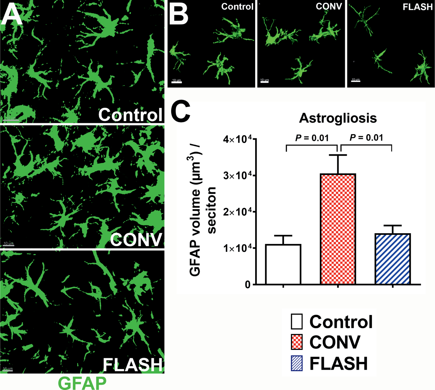
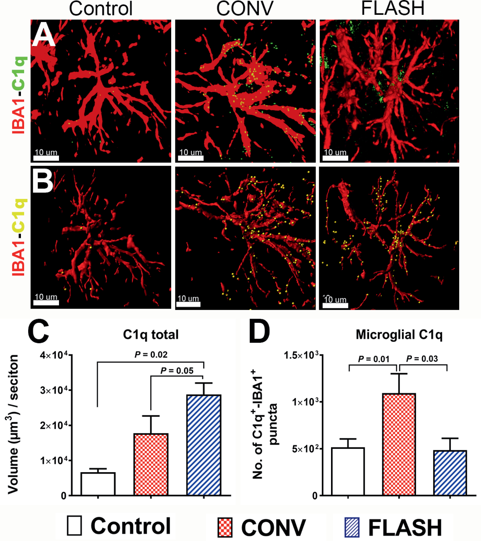
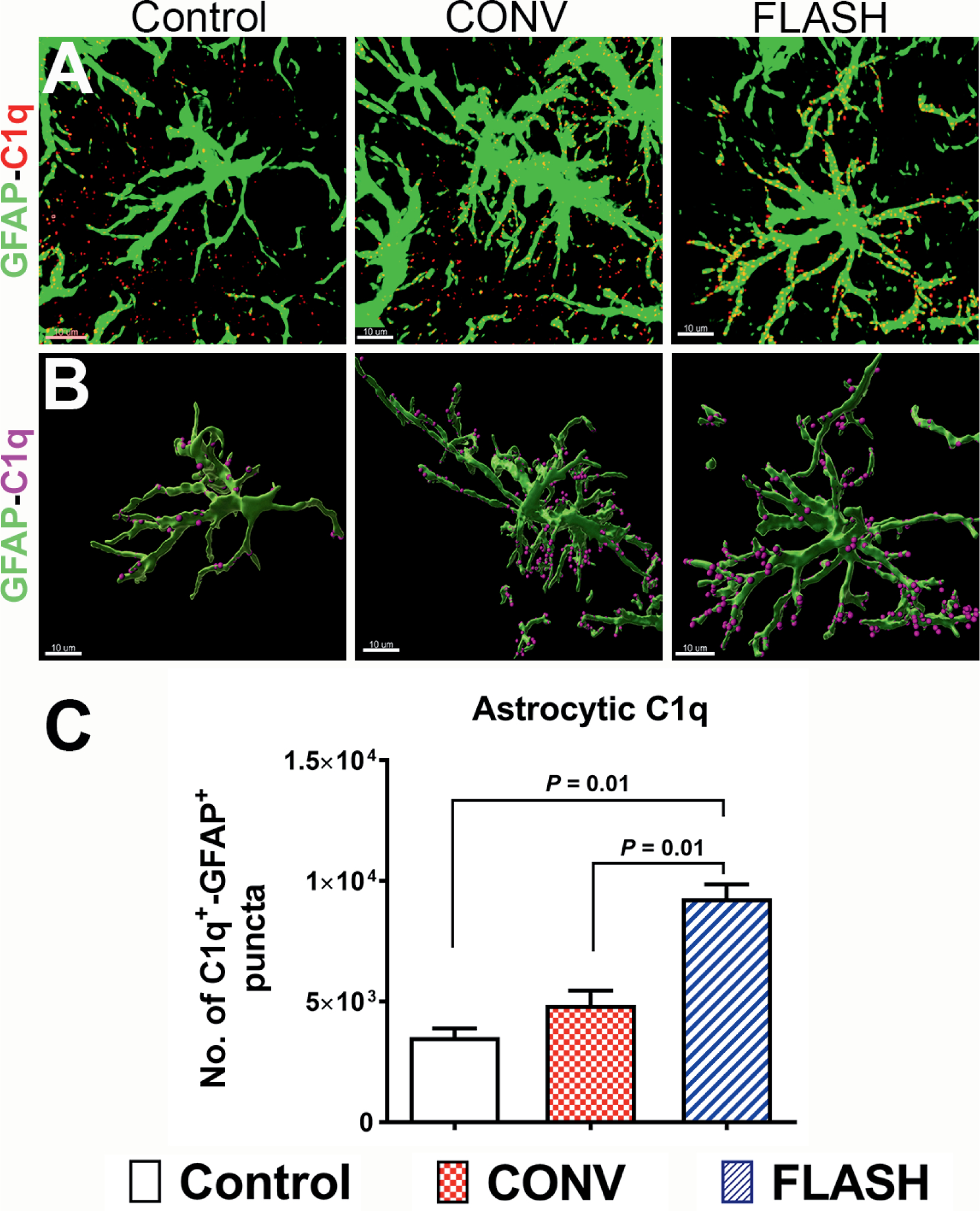
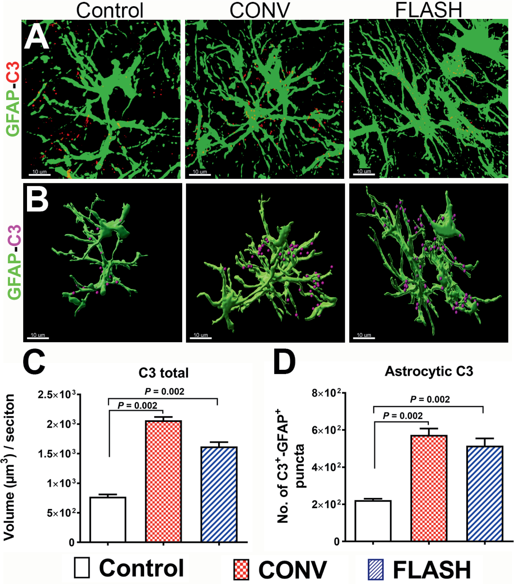
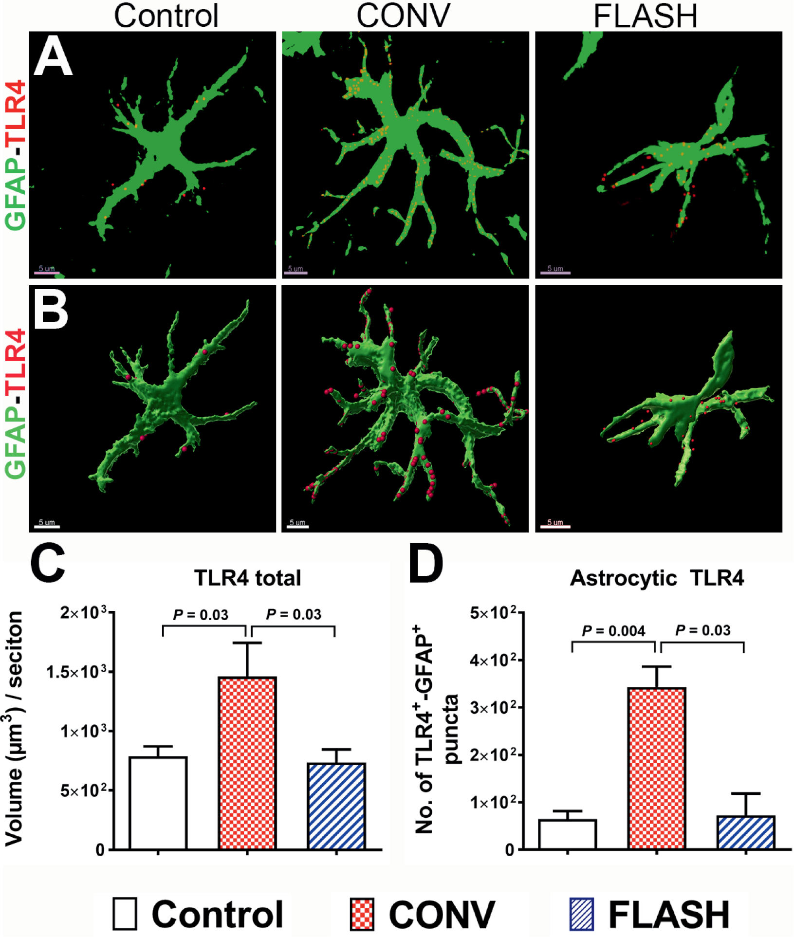
Encephalic radiation therapy delivered at a conventional dose rate (CONV, 0.1-2.0 Gy/min) elicits a variety of temporally distinct damage signatures that invariably involve persistent indications of neuroinflammation. Past work has shown an involvement of both the innate and adaptive immune systems in modulating the central nervous system (CNS) radiation injury response, where elevations in astrogliosis, microgliosis and cytokine signaling define a complex pattern of normal tissue toxicities that never completely resolve. These side effects constitute a major limitation in the management of CNS malignancies in both adult and pediatric patients. The advent of a novel ultra-high dose-rate irradiation modality termed FLASH radiotherapy (FLASH-RT, instantaneous dose rates ≥106 Gy/s; 10 Gy delivered in 1-10 pulses of 1.8 µs) has been reported to minimize a range of normal tissue toxicities typically concurrent with CONV exposures, an effect that has been coined the "FLASH effect." Since the FLASH effect has now been found to significantly limit persistent inflammatory signatures in the brain, we sought to further elucidate whether changes in astrogliosis might account for the differential dose-rate response of the irradiated brain. Here we report that markers selected for activated astrogliosis and immune signaling in the brain (glial fibrillary acidic protein, GFAP; toll-like receptor 4, TLR4) are expressed at reduced levels after FLASH irradiation compared to CONV-irradiated animals. Interestingly, while FLASH-RT did not induce astrogliosis and TLR4, the expression level of complement C1q and C3 were found to be elevated in both FLASH and CONV irradiation modalities compared to the control. Although functional outcomes in the CNS remain to be cross-validated in response to the specific changes in protein expression reported, the data provide compelling evidence that distinguishes the dose-rate response of normal tissue injury in the irradiated brain.


Figures

FIG. 1.
FIG. 1.
FLASH-RT did not induce astrocytic hypertrophy. Representative z stacks from laser scanning confocal microscopy (green, panel A) and volumetric analysis of 3D reconstruction for hippocampal astrocytes (green, GFAP, panel B) showed increased soma volume with thicker and longer stelae indicating astrocytic hypertrophy one month after 10 Gy CONV-RT. The astrocytic morphology in the FLASH-irradiated group was comparable to controls (panel C). Data are presented as mean ± SEM (n = 6 animals per group). P values are derived from non-parametric Kruskal-Wallis H test and Mann-Whitney’s comparison between each group as indicated. Scale bar = 10 μm (panels A and B).
FIG. 2.
FIG. 2.
FLASH-RT of the brain did not elevate microglial expression of complement C1q. Confocal z stacks (red, IBA1; green, C1q, panel A) and 3D algorithm-based volumetric quantification of microglia (red, panel B) co-labeled with C1q (yellow spots, panel B) showed an increased total C1q expression (panel C) in the hippocampus after both irradiation modalities (10 Gy). One month after 10 Gy CONV-RT, there was significantly elevated microglial co-labeling of C1q in the hippocampus (panel D), whereas after FLASH-RT this was not observed. Data are presented as mean ± SEM (n = 4–6 animals per group). P values are derived from non-parametric Kruskal-Wallis H test and Mann-Whitney’s comparison between each group as indicated. Scale bar = 10 μm (panels A and B).
FIG. 3.
FIG. 3.
Elevated astrocytic expression of complement C1q in the irradiated brain. Volumetric quantification of confocal z stacks (green, GFAP; red, C1q, panel A) and 3D reconstruction of GFAP+ astrocytes (green, panel B) co-labeled with C1q (magenta spots, panel B) showed a significantly elevated complement C1q one month after either 10 Gy CONV-RT or FLASH-RT (panel C). Data are presented as mean ± SEM (n = 4–6 animals per group). P values are derived from non-parametric Kruskal-Wallis H test and Mann-Whitney’s comparison between each group as indicated. Scale bar = 10 μm (panels A and B).
FIG. 4.
FIG. 4.
Cranial irradiation increased astrocytic co-labeling of complement C3 in the hippocampus. Representative z stacks from laser scanning confocal microscopy (green, GFAP; red, C3, panel A) and volumetric quantification of 3D rendered GFAP+ astrocytes (green, panel B) showed a significantly elevated total C3 and GFAP co-labeling with C3 (magenta spots, panel B) one month after either 10 Gy CONV-RT or FLASH-RT (panel C). Data are presented as mean ± SEM (n = 6 animals per group). P values are derived from non-parametric Kruskal-Wallis H test and Mann-Whitney’s comparison between each group as indicated. Scale bar = 10 μm (panel A and B).
FIG. 5.
FIG. 5.
FLASH-RT did not elevate astrocytic expression of the danger receptor TLR4. Volumetric quantification of confocal z stacks (green, GFAP; red, TLR4, panel A) and 3D reconstruction of GFAP+ surface (green, panel B) co-labeled with TLR4 (red, panel B) showed a significantly elevated total TLR4 and co-labeling with the danger-sensing receptor TLR4 one month after CONV-RT (panels C and D). Data are presented as Mean ± SEM (n = 5–6 animals per group). P values are derived from non-parametric Kruskal-Wallis H test and Mann-Whitney’s comparison between each group as indicated. Scale bar = 5 μm (panels A and B).

Similar articles

Cited by

References

    1. Vozenin MC, Hendry JH, Limoli CL. Biological benefits of ultra-high dose rate FLASH radiotherapy: Sleeping Beauty awoken. Clin Oncol (R Coll Radiol) 2019; 31:407–15. - PMC - PubMed
    1. Harrington KJ. Ultrahigh dose-rate radiotherapy: Next steps for FLASH-RT. Clin Cancer Res 2019; 25:3–5. - PubMed
    1. Montay-Gruel P, Acharya MM, Petersson K, Alikhani L, Yakkala C, Allen BD, et al. Long-term neurocognitive benefits of FLASH radiotherapy driven by reduced reactive oxygen species. Proc Natl Acad Sci U S A 2019; 116:10943–51. - PMC - PubMed
    1. Montay-Gruel P, Bouchet A, Jaccard M, Patin D, Serduc R, Aim W, et al. X-rays can trigger the FLASH effect: Ultra-high dose-rate synchrotron light source prevents normal brain injury after whole brain irradiation in mice. Radiother Oncol 2018; 129:582–8. - PubMed
    1. Montay-Gruel P, Petersson K, Jaccard M, Boivin G, Germond JF, Petit B, et al. Irradiation in a flash: Unique sparing of memory in mice after whole brain irradiation with dose rates above 100 Gy/s. Radiother Oncol 2017; 124:365–9. - PubMed

Publication types