Case Reports
. 2021 Jan;28(1):248-258.
doi: 10.1111/ene.14491. Epub 2020 Oct 5.

Cytokine release syndrome-associated encephalopathy in patients with COVID-19

Affiliations
Case Reports

Cytokine release syndrome-associated encephalopathy in patients with COVID-19

P Perrin et al. Eur J Neurol. 2021 Jan.

Abstract

Background and purpose: Neurological manifestations in coronavirus disease (COVID)-2019 may adversely affect clinical outcomes. Severe COVID-19 and uremia are risk factors for neurological complications. However, the lack of insight into their pathogenesis, particularly with respect to the role of the cytokine release syndrome (CRS), is currently hampering effective therapeutic interventions. The aims of this study were to describe the neurological manifestations of patients with COVID-19 and to gain pathophysiological insights with respect to CRS.

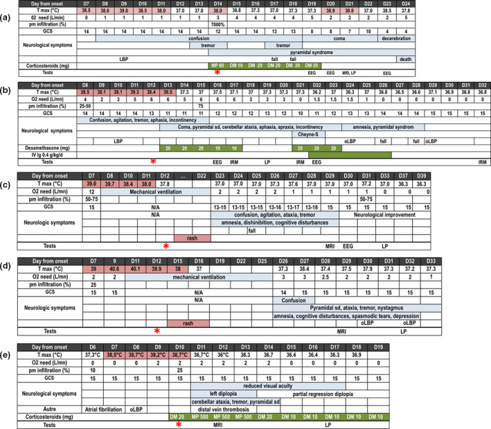
Methods: In this longitudinal study, we performed extensive clinical, laboratory and imaging phenotyping in five patients admitted to our renal unit.

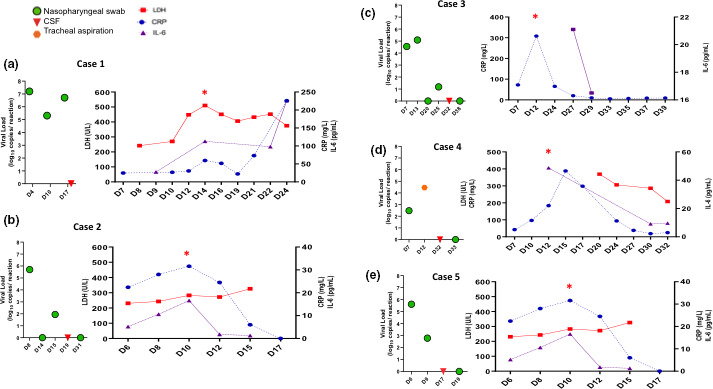
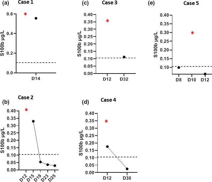
Results: Neurological presentation included confusion, tremor, cerebellar ataxia, behavioral alterations, aphasia, pyramidal syndrome, coma, cranial nerve palsy, dysautonomia, and central hypothyroidism. Notably, neurological disturbances were accompanied by laboratory evidence of CRS. Severe acute respiratory syndrome coronavirus-2 (SARS-CoV-2) was undetectable in the cerebrospinal fluid (CSF). Hyperalbuminorrachia and increased levels of the astroglial protein S100B were suggestive of blood-brain barrier (BBB) dysfunction. Brain magnetic resonance imaging findings comprised evidence of acute leukoencephalitis (n = 3, one of whom had a hemorrhagic form), cytotoxic edema mimicking ischaemic stroke (n = 1), or normal results (n = 2). Treatment with corticosteroids and/or intravenous immunoglobulins was attempted, resulting in rapid recovery from neurological disturbances in two cases. SARS-CoV2 was undetectable in 88 of the 90 patients with COVID-19 who underwent Reverse Transcription-PCR testing of CSF.

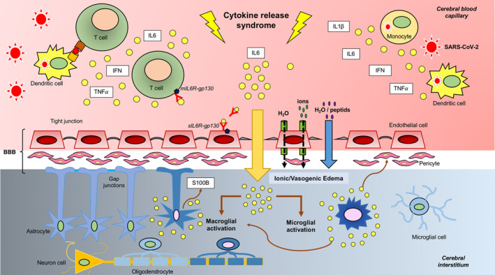
Conclusions: Patients with COVID-19 can develop neurological manifestations that share clinical, laboratory and imaging similarities with those of chimeric antigen receptor T-cell-related encephalopathy. The pathophysiological underpinnings appear to involve CRS, endothelial activation, BBB dysfunction, and immune-mediated mechanisms.

Keywords: COVID-19; corticosteroids; cytokine; encephalitis; intravenous immunoglobulins; kidney; neurological disorders.


Conflict of interest statement

The authors declare no financial or other conflicts of interest.

Figures

Figure 1
Figure 1
Panels a−e. Temporal course of clinical and laboratory variables in the five patients studied. Diagnostic investigations and administered drugs are also reported. All longitudinal data are shown with respect to the date of COVID‐19 symptom onset (D0). Red stars denote the days on which levels of cytokine release syndrome‐related inflammatory biomarkers reached a peak. % pm infiltration, percentage of lung infiltration on chest scan; CSF, cerebrospinal fluid; DM, dexamethasone; EEG, electroencephalogram; GCS, Glasgow coma scale; LBP, low blood pressure; MP, methylprednisolone; oLBP, orthostatic hypotension; sd, syndrome; Tmax, maximum body temperature; N/A, not available, VA, visual acuity. [Colour figure can be viewed at wileyonlinelibrary.com]
Figure 2
Figure 2
Brain magnetic resonance imaging (MRI) findings in the five patients studied. Case 1: Axial fluid‐attenuated inversion recovery (FLAIR)‐weighted MRI (a), axial diffusion‐weighted MRI (b) and axial apparent diffusion coefficient (ADC) map (c). MRI images obtained on day 21; cross‐sections through motor areas, frontal and parietal lobes. Diffuse bilateral, symmetric white matter FLAIR hyperintensities with mild hyperintense 'ground glass' areas (arrow heads) and frank hyperintense areas (conventional radiological FLAIR hyperintensities; arrows). All abnormal FLAIR areas appeared hyperintense in the diffusion sequence and were characterized by a gradient: frank hyperintense FLAIR lesions had a higher intensity than 'ground glass' areas. On the ADC map, 'ground glass' areas were iso‐ or hypointense, whereas frank hyperintense FLAIR areas were hyperintense. Abnormal FLAIR hyperintensities were preferentially localized to subcortical white matter of motor areas and showed a bilateral symmetric distribution. The anterior frontal white matter appeared normal on FLAIR and diffusion‐weighted imaging (DWI) sequences (a, b: stars). Case 2: Axial DWI (d) and axial ADC map (e): cross‐sections through frontal and parietal lobes on day 17. Axial DWI (f) and axial ADC map (g): cross‐sections through frontal and parietal lobes on D33. Coronal FLAIR weighted MRI on day 33 (h): cross‐section through middle cerebellar peduncles. The first MRI (on D17) revealed an acute hyperintense DWI lesion, with hypointensity on ADC map, suggestive of a cytotoxic edema in the left frontal lobe (d, e) and deemed initially compatible with an acute stroke. However, this lesion maintained a similar aspect following 16 days (f, g), casting doubts on its ischaemic origin. Persistence of middle cerebellar peduncles hyperintensities was also evident (h). Case 4: Axial susceptibility‐weighted imaging MRI at day 31: cross‐section through motor areas, frontal and parietal lobes (i) and the splenium (j). Axial FLAIR weighted MRI (k): cross‐section through the splenium. Multifocal microbleeds were evident in the splenium (j: black arrow heads) and in the white matter/gray matter junction (i: black arrow head), with an apparent perivascular distribution in the Virchow‐Robin spaces. These lesions were associated with hyperintensities on FLAIR weighted sequence (k: arrows). Day 0 = date of COVID‐19 symptom onset.
Figure 3
Figure 3
Temporal course of viral loads and biomarkers of cytokine release syndrome (CRS) in the five study patients. Red stars denote the days on which levels of CRS‐related inflammatory biomarkers reached a peak. Reference values: interleukin (IL)‐6, <4 ng/l; lactate dehydrogenase (LDH), 120−246 UI/l; C‐reactive protein (CRP) <4 mg/l. All longitudinal data are shown with respect to the date of COVID‐19 symptom onset (D0). [Colour figure can be viewed at wileyonlinelibrary.com]
Figure 4
Figure 4
Temporal course of serum S100B (µg/l) levels in the five patients studied. Values above the dotted line indicate high values (>0.105 µg/l). Red stars denote the days on which levels of cytokine release syndrome‐related inflammatory biomarkers reached a peak. All longitudinal data are shown with respect to the date of COVID‐19 symptom onset (D0). [Colour figure can be viewed at wileyonlinelibrary.com]
Figure 5
Figure 5
Cytokine release syndrome (CRS)‐associated encephalitis in COVID‐19: pathophysiological model. In severe cases of COVID‐19, SARS‐CoV‐2 induces CRS during the second week of infection. Following viral infection, macrophages, dendritic cells, other immune cells and endothelial cells become activated and produce large amounts of proinflammatory molecules [including interleukin (IL)‐6, IL‐1β, tumor necrosis factor (TNF)‐α, interferon (IFN)‐α/β, and IFN‐γ]. IL‐6 acts as a master mediator of a self‐perpetuating proinflammatory loop that results in lymphocyte activation (via the cis‐pathway) followed by a massive release of cytokines (cytokine storm). The trans‐pathway of activation may alter endothelial permeability, resulting in blood−brain barrier (BBB) dysfunction [20, 27]. In turn, an altered BBB permeability may lead to edema and even red blood cells extravasation (potentially accounting for the hemorrhagic form of acute leukoencephalitis observed in case #4). This sequence of events closely resembles those occurring in immune effector cell neurotoxicity syndrome [18]. Proinflammatory cytokines can leak through a dysfunctional BBB, ultimately activating microglial cells (brain tissue‐resident macrophages). This may, in turn, lead to a secondary inflammatory response in the macroglia accompanied by the release of S100B, an astroglial protein reflecting both glial activation and BBB dysfunction. The resulting neuroinflammatory response can yield to reactive gliosis accompanied by infiltration of CD68+ monocytes/macrophage [21, 22]. ; gp130, glycoprotein 130; sIL6R, soluble interleukin‐6 receptor. [Colour figure can be viewed at wileyonlinelibrary.com]

References

    1. Mao L, Jin H, Wang M, et al. Neurologic manifestations of hospitalized patients with coronavirus disease 2019 in Wuhan, China. JAMA Neurol 2019; 77: 1–9. - PMC - PubMed
    1. Helms J, Kremer S, Merdji H, et al. Neurologic features in severe SARS‐CoV‐2 infection. N Engl J Med 2020; 382: 2268–2270. - PMC - PubMed
    1. Romoli M, Jelcic I, Bernard‐Valnet R, et al. The infectious disease panel of the European academy of neurology a systematic review of neurological manifestations of SARS‐CoV‐2 infection: the devil is hidden in the details. Eur J Neurol 2020; 27: 1712–1726. - PMC - PubMed
    1. Wu Y, Xu X, Chen Z, et al. Nervous system involvement after infection with COVID‐19 and other coronaviruses. Brain Behav Immun 2020; 87: 18–22. - PMC - PubMed
    1. Mehta P, McAuley DF, Brown M, Sanchez E, Tattersall RS, Manson JJ. HLH across speciality collaboration, UK COVID‐19: consider cytokine storm syndromes and immunosuppression. Lancet 2020; 395: 1033–1034. - PMC - PubMed

Publication types

MeSH terms