Review
. 2020 Sep 3;79(5):710-727.
doi: 10.1016/j.molcel.2020.07.027. Epub 2020 Aug 4.

Coronavirus RNA Proofreading: Molecular Basis and Therapeutic Targeting

Affiliations
Review

Coronavirus RNA Proofreading: Molecular Basis and Therapeutic Targeting

Fran Robson et al. Mol Cell. .

Erratum in

Abstract

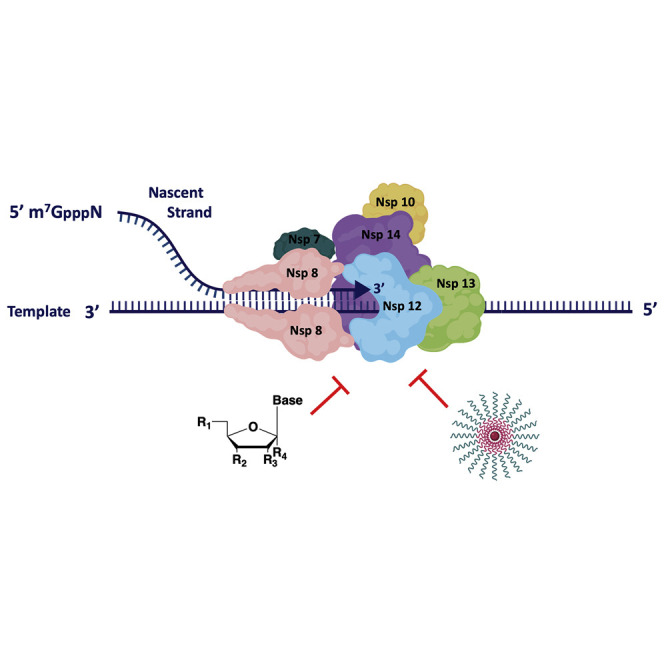
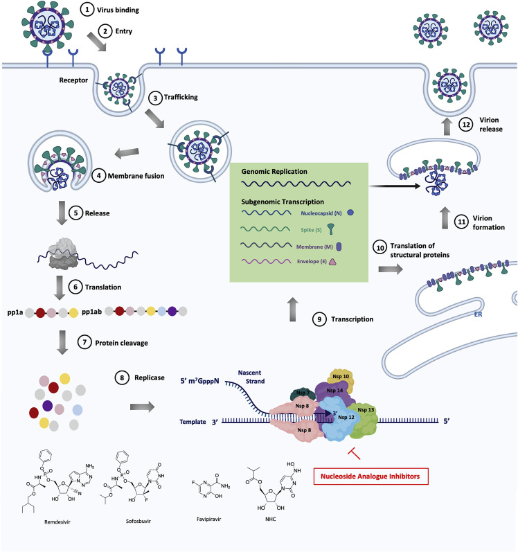
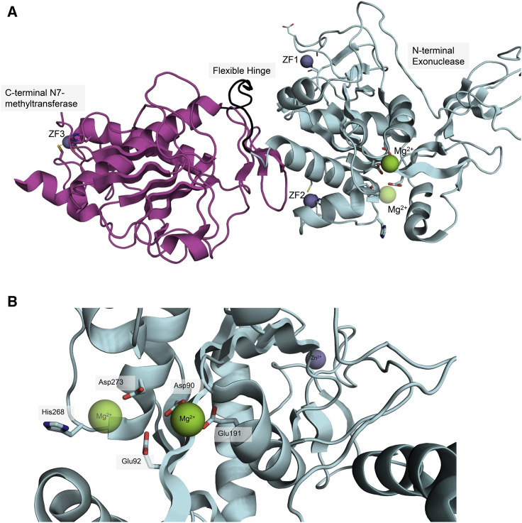
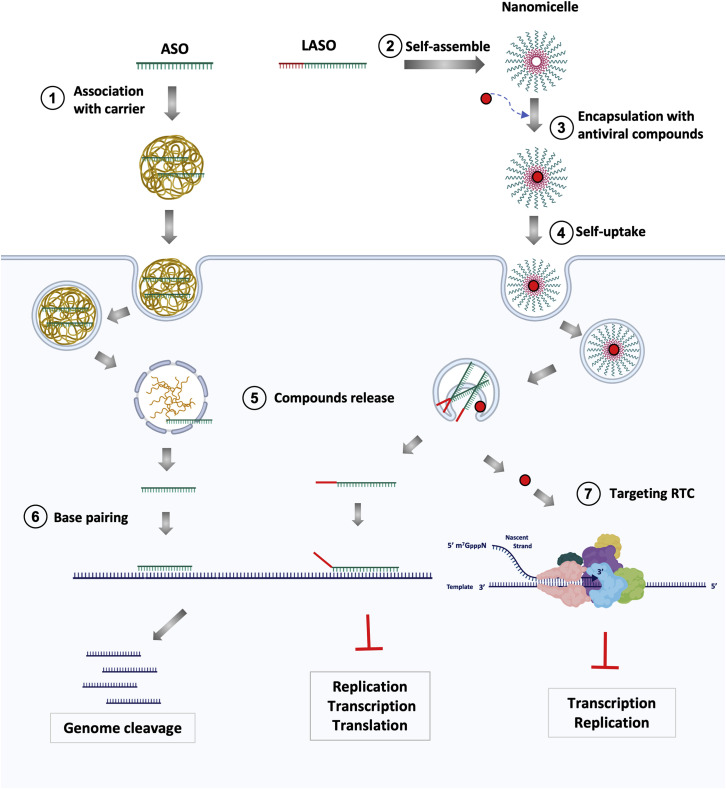
The coronavirus disease 2019 (COVID-19) that is wreaking havoc on worldwide public health and economies has heightened awareness about the lack of effective antiviral treatments for human coronaviruses (CoVs). Many current antivirals, notably nucleoside analogs (NAs), exert their effect by incorporation into viral genomes and subsequent disruption of viral replication and fidelity. The development of anti-CoV drugs has long been hindered by the capacity of CoVs to proofread and remove mismatched nucleotides during genome replication and transcription. Here, we review the molecular basis of the CoV proofreading complex and evaluate its potential as a drug target. We also consider existing nucleoside analogs and novel genomic techniques as potential anti-CoV therapeutics that could be used individually or in combination to target the proofreading mechanism.

Keywords: ASO; CoV; ExoN; NA; anti-coronavirus drugs; antisense oligonucleotide; coronavirus; exonuclease; non-structural protein 14; nsp14; nucleoside analog.


Conflict of interest statement

Declaration of Interests The authors declare no competing interests.

Figures

None
Graphical abstract
Figure 1
Figure 1
Single-Stranded RNA Genome of SARS-CoV-2 Two-thirds of the genome encodes two large polyproteins, pp1a and pp1ab, that are cleaved into 16 non-structural proteins. The last one-third of the genome encodes structural and accessory proteins. This figure was created with BioRender.
Figure 2
Figure 2
Virus Replication Mechanism To enter in the host cell, first the virus binds to the ACE2 receptor (1) to initiate the viral entry (2), the vacuole containing the virus is then internalized (3) and the membrane fuses with the virus (4) in order to release it (5) into the cytoplasm of host cell. The genome is then translated to produce the polyproteins pp1a and pp1ab (6), which are cleaved by proteases (7) to yield the 16 NSPs that form the RNA replicase-transcriptase complex (8). Viral genome is duplicated and mRNA encoding structural proteins are transcribed (9). Then, the subgenomic mRNAs are translated into structural proteins (10). The formation of the new virion takes place on modified intracellular membranes that are derived from the rough endoplasmic reticulum (ER) in the perinuclear region (11). The new virion is then released (12). In red are localized the sites of action of a number of small-molecule antivirals. This figure was created with BioRender.
Figure 3
Figure 3
Model of the Core Replication and Proofreading Complex of SARS-CoV Nsp12-RdRp replicates and transcribes the genome and sgmRNAs. Nsp7/nsp8 proteins confer processivity to the polymerase. Nsp13 unwinds dsRNA ahead of the polymerase. Nsp14-ExoN complexed with its co-factor nsp10 proofreads the nascent RNA strand and excises misincorporated nucleotides. Nsp13, an unknown GTPase, Nsp14-N7-methyltransferase, and the Nsp16-2′-O-methyltransferase/Nsp10 complex are involved in the capping mechanism. This figure was created with BioRender.
Figure 4
Figure 4
The Overall Structure of Nonstructural Protein 14 (A) Cartoon representation of the structure of the nsp14. The N-terminal exonuclease domain (aquamarine) and C-terminal N7-methyltransferase (light magenta) are connected by a flexible interdomain loop (black). The amino acid residues that coordinate zinc fingers (ZF) (slate blue) and magnesium cofactor (Mg2+) (pea green) are shown as sticks. Protein structure is retrieved from Protein Data Bank (PDB ID: 5C8U; Ma et al., 2015). (B) Detailed cartoon representation of the catalytic DEEDh residues. DEEDh domain comprises Asp90, Glu92, Glu191, Asp273, and His268 amino acids residues that are located in the exonuclease domain (aquamarine) of the nsp14. Mg2+ cofactor (pea green) is coordinated by Asp90 and Glu191 and is thought to facilitate the removal of misincorporated nucleotides. The second Mg2+ is shown tentatively; isothermal titration calorimetry predicted a two-metal binding mode for divalent cations, but crystallography data showed only one (Chen et al., 2007). The protein structure was retrieved from Protein Data Bank (PDB: 5C8U; Ma et al., 2015).
Figure 5
Figure 5
ASO Technology in Therapy against SARS-CoV-2 Antisense oligonucleotides (ASOs) are conjugated with a carrier that allows delivery into the cells (1). Lipid-modified ASOs (LASO) self-assemble into nanomicelles (2) and encapsulate an antiviral molecule such as an NA (3). These nanomicelles are able to enter the cell without any transfection agents (4). Once inside the cell, ASO, LASO, and the encapsulated drugs are released (5). ASOs are administered via cationic polymers and are released through the proton sponge effect (the presence of the weakly basic molecule causes the endosome to burst). LASOs enter via the macropinocytosis mechanism and are released through interaction with the endosomal membrane. The intracellular ASOs/LASOs match to their complementary sequences (6), leading either to genome degradation (through RNaseH activity) or replication/transcription/translation blocks due to steric block formation. The NAs can interfere with the RNA replication and transcription by targeting RdRp as described above (7). This figure was created with BioRender.

Similar articles

Cited by

References

    1. Abbott T.R., Dhamdhere G., Liu Y., Lin X., Goudy L., Zeng L., Chemparathy A., Chmura S., Heaton N.S., Debs R., et al. Development of CRISPR as an Antiviral Strategy to Combat SARS-CoV-2 and Influenza. Cell. 2020;181:865–876. - PMC - PubMed
    1. Adedeji A.O., Marchand B., Te Velthuis A.J.W., Snijder E.J., Weiss S., Eoff R.L., Singh K., Sarafianos S.G. Mechanism of nucleic acid unwinding by SARS-CoV helicase. PLoS ONE. 2012;7:e36521. - PMC - PubMed
    1. Agostini M.L., Andres E.L., Sims A.C., Graham R.L., Sheahan T.P., Lu X., Smith E.C., Case J.B., Feng J.Y., Jordan R., et al. Coronavirus Susceptibility to the Antiviral Remdesivir (GS-5734) Is Mediated by the Viral Polymerase and the Proofreading Exoribonuclease. MBio. 2018;9 doi: 10.1128/mBio.00221-18. Published online March 6, 2018. - DOI - PMC - PubMed
    1. Agostini M.L., Pruijssers A.J., Chappell J.D., Gribble J., Lu X., Andres E.L., Bluemling G.R., Lockwood M.A., Sheahan T.P., Sims A.C., et al. Small-Molecule Antiviral β-d-N4-Hydroxycytidine Inhibits a Proofreading-Intact Coronavirus with a High Genetic Barrier to Resistance. J. Virol. 2019;93 doi: 10.1128/JVI.01348-19. Published online November 26, 2019. - DOI - PMC - PubMed
    1. Almeida J.D., Berry D.M., Cunningham C.H., Hamre D., Hofstad M.S., Mallucci L., McIntosh K., Tyrrell D.A.J. Virology: Coronaviruses. Nature. 1968;220:650. 650.

Publication types

MeSH terms