. 2020 Nov:95:102946.
doi: 10.1016/j.dnarep.2020.102946. Epub 2020 Aug 15.

C17orf53 is identified as a novel gene involved in inter-strand crosslink repair

Affiliations

C17orf53 is identified as a novel gene involved in inter-strand crosslink repair

Chao Wang et al. DNA Repair (Amst). 2020 Nov.

Abstract

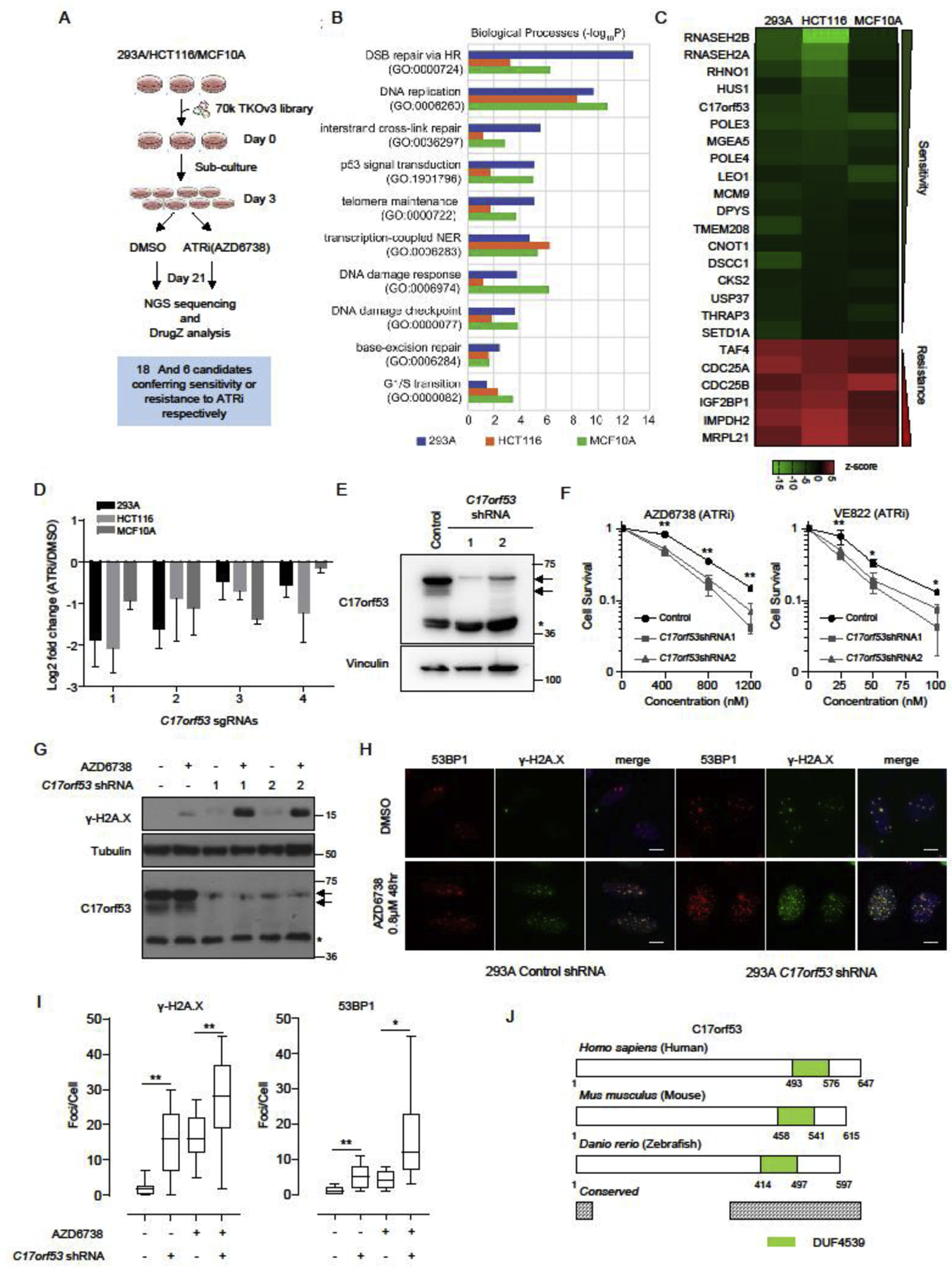
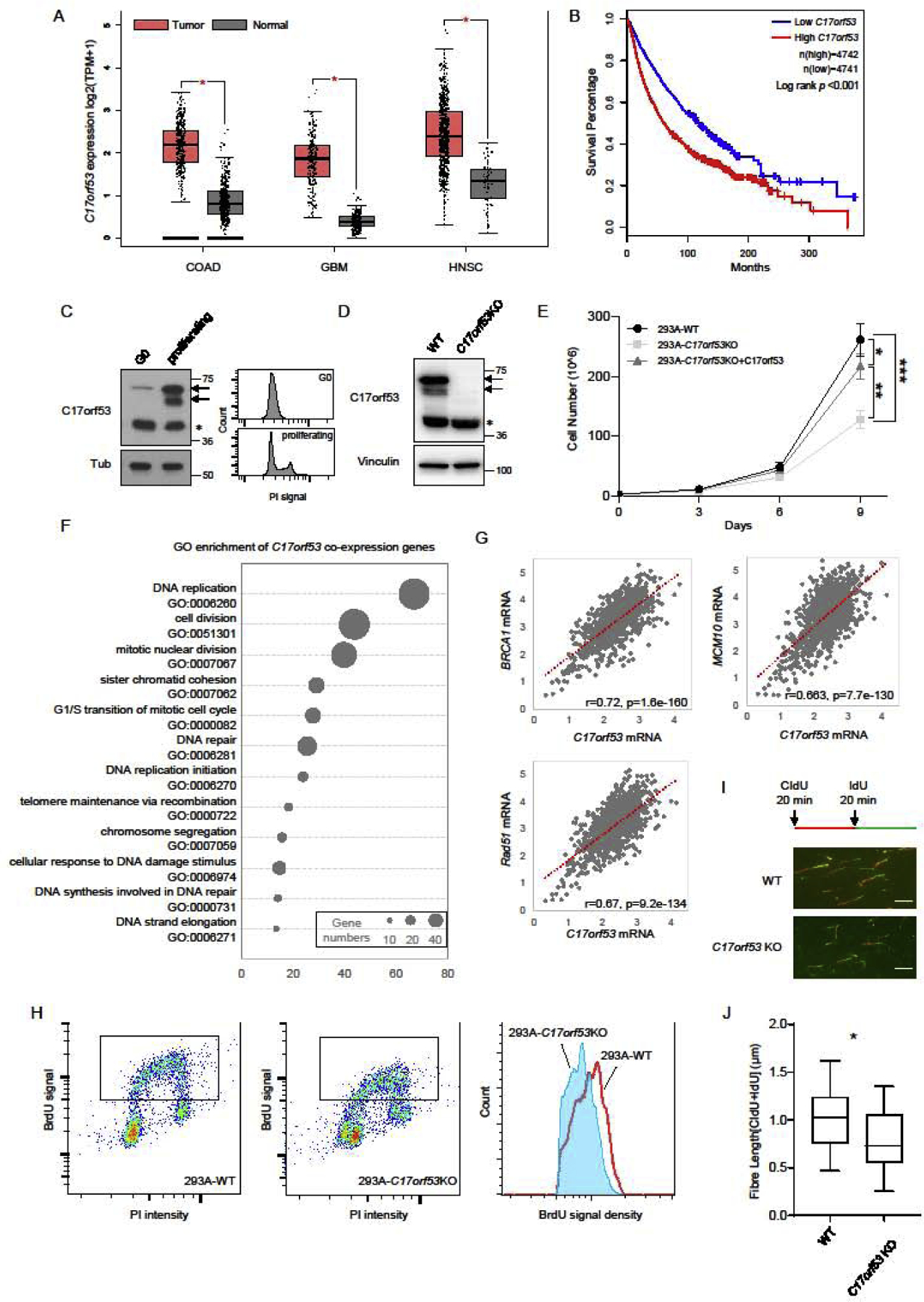
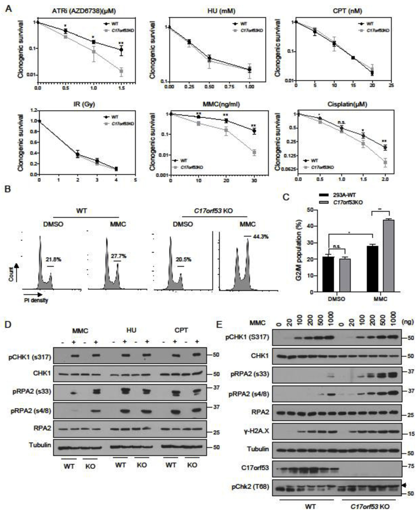
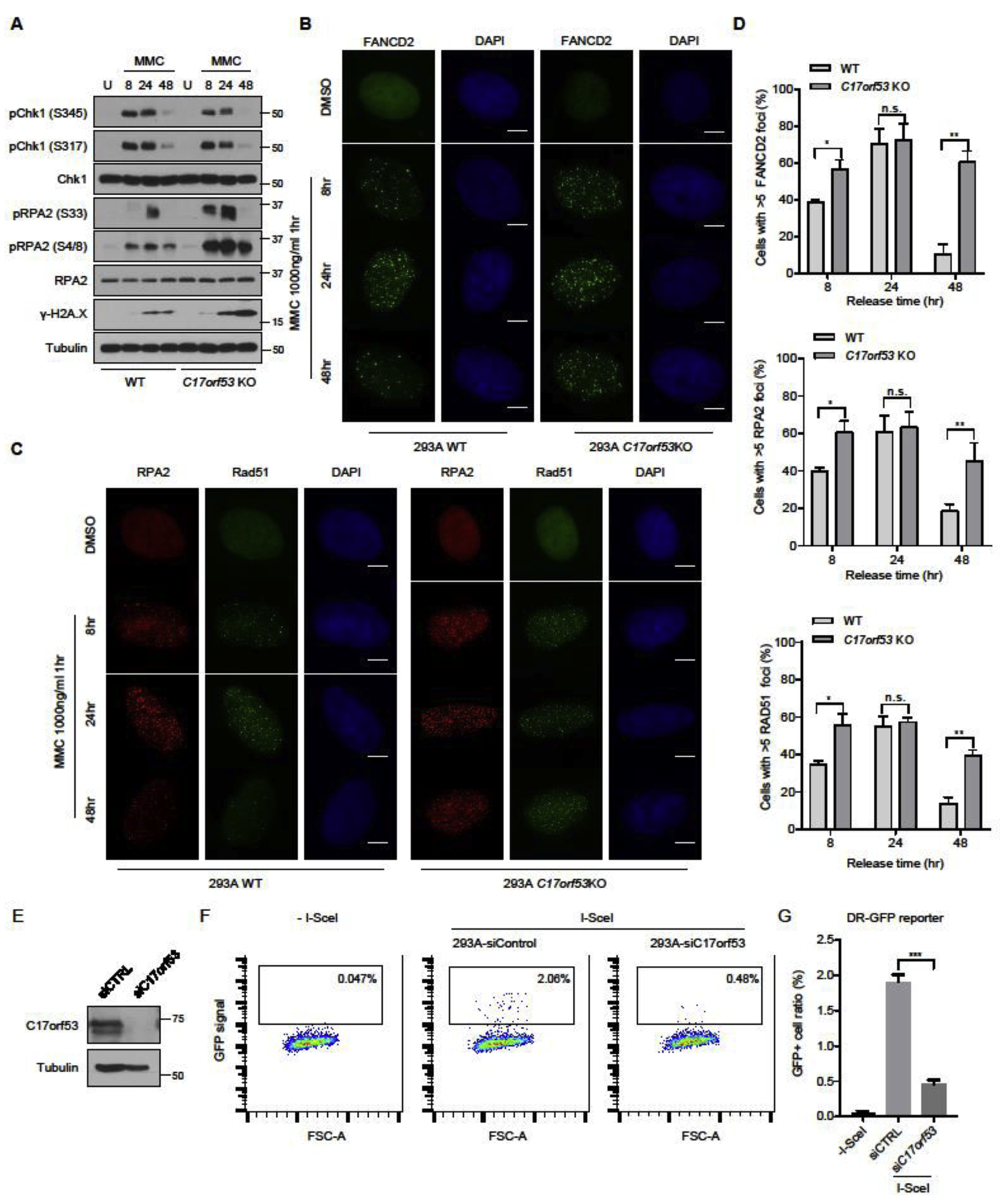
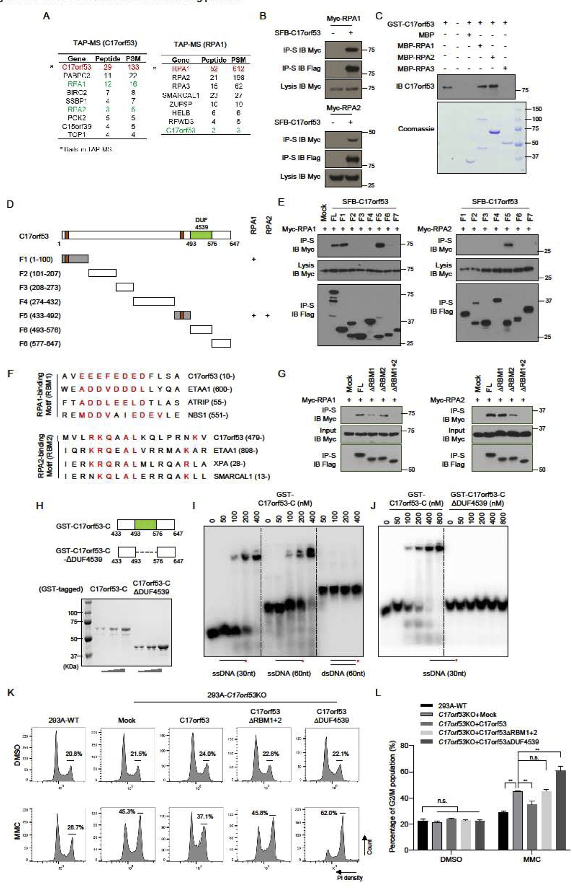
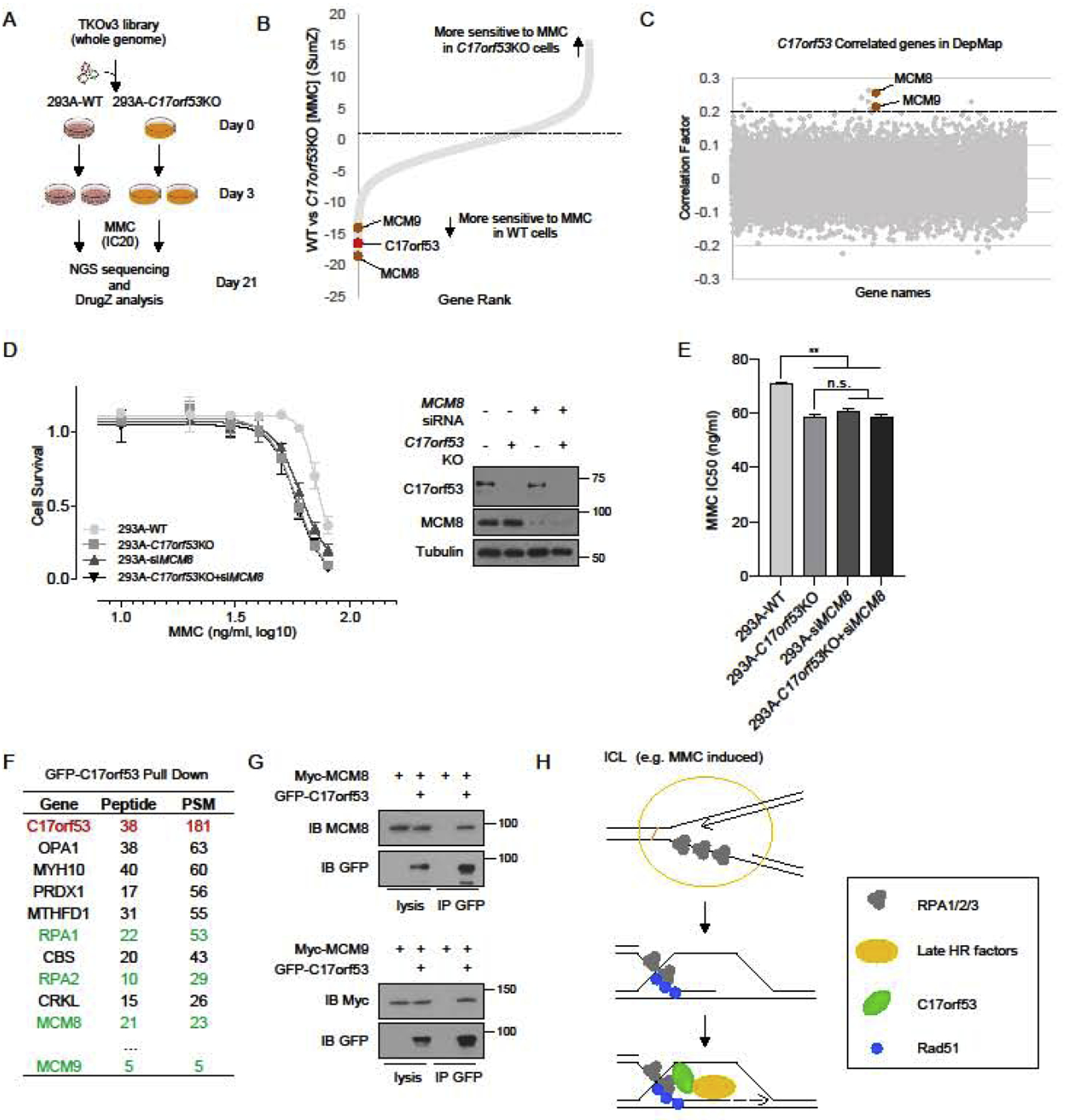
Ataxia Telangiectasia and Rad3-Related kinase (ATR) is a master regulator of genome maintenance, and participates in DNA replication and various DNA repair pathways. In a genome-wide screen for ATR-dependent fitness genes, we identified a previously uncharacterized gene, C17orf53, whose loss led to hypersensitivity to ATR inhibition. C17orf53 is conserved in vertebrates and is required for efficient cell proliferation. Loss of C17orf53 slowed down DNA replication and led to pronounced interstrand crosslink (ICL) repair defect. We showed that C17orf53 is a ssDNA- and RPA-binding protein and both characteristics are important for its functions in the cell. In addition, using multiple omics methods, we found that C17orf53 works with MCM8/9 to promote cell survival in response to ICL lesions. Taken together, our data suggest that C17orf53 is a novel component involved in ICL repair pathway.

Keywords: C17orf53; DNA replication; Genome-wide screen; ICL repair; RPA-binding, HROB, MCM8IP; ssDNA-binding.


Conflict of interest statement

Conflict of interest statement

The authors declare no competing financial interests.

Figures

Figure 1.
Figure 1.. C17orf53 was Identified as a Determinant of ATRi Sensitivity.
(A) Schematic representation of the workflow for CRISPR screens performed in 293A, HCT116, and MCF10A cells. (B) Gene ontology (GO) analysis of the synthetic lethality genes identified in the CRISPR screens. The top10 GO terms significantly enriched are listed here. Different color represents different cell line. Blue: 293A, Orange: HCT116, Green: MCF10A. (C) Visualization of the high-confidence genes in these screens. The drugZ scores are displayed as a heatmap summarizing the common genes uncovered in all three screens (FDR<0.01) which show synthetic lethality (score<0) or synthetic survival (score>0) with ATR inhibition. (D) Fold change ratios of sgRNAs targeting C17orf53 in 3 cell lines comparing ATRi treated group with DMSO treated group. Mean±SD, n=3. (E) Immunoblot of wild-type and C17orf53 knock-down 293A cells. Arrows mark the C17orf53 protein, asterisk marks a non-specific band. (F) Clonogenic survival assay of cells treated with ATR inhibitors AZD6738 or VE-822. Mean±SD, n=3, *p<0.05, **p<0.01, student t-test. (G) Wild-type and C17orf53 knock-down 293A cells were treated with AZD6738 (0.8 μM, 48hr). Cells were harvested and lysates were analyzed by immunoblotting with indicated antibodies. Arrows mark the C17orf53 protein, asterisk marks a non-specific band. (H) Wild-type and C17orf53 knock-down 293A cells were treated with DMSO or AZD6738 (0.8 μM, 48hr). Cells were fixed and processed for γ-H2A.X and 53BP1 immunofluorescence staining. The scale bar represents 1 μm. (I) Quantification of γ-H2A.X and 53BP1 foci in different groups in H). n=100 cells, *P<0.05, **P<0.01, student t-test. (J) Schematic diagram of C17orf53 proteins from human to zebrafish. Green color marks a conserved domain which was named as Domain with Unknown Function 4539 (DUF4539). The protein regions with >80% sequence similarity from zebrafish to human were marked as “conserved” (Grey).
Figure 2.
Figure 2.. C17orf53 is Necessary for Optimal Cell Proliferation and DNA Replication.
(A) C17orf53 expression is upregulated in various types of tumors (data from TCGA). Analysis of C17orf53 expression (log2 [TPM+1]) in TCGA tumors and the matched normal tissues. COAD: Colon adenocarcinoma; GBM: Glioblastoma; HNSC: Head and Neck squamous cell carcinoma. *p<0.01, Wilcoxon test. (B) Kaplan-Meier curves of overall survival for the overall population (more than 9000, data from TCGA) according to the expression levels of C17orf53. Statistical significance was assessed by the log-rank test and presented. (C) 293A cells were contact inhibited (G0) then released for 12 hrs (proliferating). (Left panel) Total cell lysates were prepared from each group and immunoblotted with the indicated antibodies. Asterisk marks the non-specific band. (Right Panel) The same samples were fixed, stained with Propidium iodide (PI) and analyzed by fluorescence-activated cell sorting (FACS). (D) Immunoblot analysis of 293A wild-type and C17orf53 KO cells. Arrows mark the C17orf53 protein, asterisk marks non-specific band. (E) Growth curves of indicated cells. Different cells were plated in 6-well plates, passaged and counted every 3 days. *p<0.05, **p<0.01, ***p<0.001, ANOVA test. (F) Gene ontology (GO) terms significant enriched among the genes co-expressed with C17orf53 (Gene list is available in Table S1). (G) Expression level of C17orf53 correlates with expression levels of BRCA1, RAD51 and MCM10 in 1019 cell lines in CCLE. Expression level was calculated by log2.FPKM+1. The correlation scores were measured by Pearson correlation. (H) BrdU incorporation assay. Growing wild-type and C17orf53KO 293A cells were incubated in medium containing BrdU (10 μM) for 30 mins, fixed, denatured with 2N HCl and stained with BrdU antibody and propidium iodide (PI). Cells with BrdU incorporation are marked with frame and histograms of BrdU intensity are shown in the right panel. (I) Quantitation of the flow cytometry data shown in (H), presented as the mean BrdU fluorescence intensity. (±SD, n=3, *p<0.05). (J) 293A WT and C17orf53 KO were treated with CIdU (25 μM) for 20 mins and then treated with IdU (250 μM) for 20 mins. DNA fibers from these cells are indicated. The scale bar represents 1 μm. (K) Quantification of DNA fiber lengths in J). Both CIdU and IdU fibers were counted. The box plots show twenty-fifth to seventy-fifth percentiles with lines indicating the median. n =100, *p<0.05, student t-test.
Figure 3.
Figure 3.. C17orf53 Deficient Cells Show Pronounced ICL Repair Response Defects.
(A) Clonogenic survival assay for WT and C17orf53KO 293A cells exposed to ATRi (AZD6738), HU, CPT, IR, MMC and Cisplatin. Mean±SD, n=3, *p<0.05, **p<0.01, student t-test. (B) 293A WT or C17orf53 KO cells were treated with DMSO or MMC (10 ng/ml) for 48 hr before ethanol fixation, propidium iodide (PI) staining, and fluorescence-activated cell sorting (FACS) analysis. Cells in G2-M cell cycle stages were marked and measured. Representative data are shown. (C) Quantification of the experiments shown in B). Mean±SD, n=3, *p<0.05, **p<0.01, student t-test. (D) WT or C17orf53KO 293A cells were treated with MMC (500 ng/ml), HU (4 mM) or CPT (200 nM) for 8 hours and then lysed. The total cell lysates were immunoblotted with the indicated antibodies. (E) WT or C17orf53KO 293A cells were treated with MMC of different doses for 8 hours and then lysed. The total cell lysates were immunoblotted with the indicated antibodies.
Figure 4.
Figure 4.. C17orf53 participates in ICL repair through affecting HR repair.
(A) WT cells and C17orf53 KO cells were treated with DMSO or MMC (1000 ng/ml, 1 hr). The cells were harvested at different time points after treatment. The total cell lysates were immunoblotted with the indicated antibodies. (B) MMC-induced FANCD2 foci formation in WT and C17orf53 KO 293A cells. The indicated cells were left untreated or treated with MMC (1000 ng/ml, 1hrs). At different time points after treatment, the cells were fixed and immunostained with FANCD2 antibody. The scale bar represents 1 μm. (C) MMC-induced RPA2 and RAD51 foci in WT and C17orf53KO 293A cells. The same cells in B) were fixed and immunostained with RPA2 and RAD51 antibodies. The scale bar represents 1 μm. (D) Quantification of FANCD2, RPA2 and RAD51 FANCD2 foci in B) and C). n=3 biological independent experiments. *p<0.05, **p<0.01, student t-test. (E) U2OS cells were transfected with control siRNA or C17orf53 siRNA. Cell lysates were immunoblotted with indicated antibodies. (F) Loss of C17orf53 results in decreased HR-directed DNA repair. U2OS-DR-GFP were transfected with control siRNA or C17orf53 siRNA, followed by transfection of I-SceI. 48hr after transfection, cells were harvested and assayed for GFP expression by FACS. Representative data are shown. (G) Quantification of the experiments shown in F). Mean±SD, n=3. ***p<0.001, student t-test.
Figure 5.
Figure 5.. C17orf53 is a RPA-binding Protein and Contains a ssDNA-binding Domain DUF4539
(A) 293T cells stably expressing SFB-C17orf53 or SFB-RPA1 were used for TAP. Tables are representative proteins identified by mass spectrometry analysis. Letters in Red indicate the bait proteins. Letters in Green indicate the candidate proteins. (B) C17orf53 interacts with RPA1 and RPA2. 293T cells were transiently transfected with indicated plasmids. Cell lysates were immunoprecipitated with S beads, and immunoblot was performed with the indicated antibodies. (C) Direct binding between recombinant GST-C17orf53 and MBP-tagged RPA1 and RPA2. Upper panel: C17orf53 was detected by immunoblotting. Lower panel: purified proteins were visualized by Commassie staining. Due to its low concentration, GST-C17orf53 protein could only be detected by immunoblotting. (D) Schematic diagram of C17orf53 indicating the truncation fragments tested in the following experiments. + represents the positive binding. (E) 293T cells were transiently transfected with indicated plasmids. Cell lysates were immunoprecipitated with S beads, and immunoblot was performed with the indicated antibodies. (F) The protein sequence of C17orf53 was compared with the sequences of known RPA-binding proteins and the similar motifs were listed above. They were named as RBM1 (RPA1-binding Motif) and RBM2 (RPA2-binding Motif). (G) 293T cells were transiently transfected with indicated plasmids. Cell lysates were immunoprecipitated with S beads, and immunoblot was performed with the indicated antibodies. (H) Schematic diagram of C17orf53 indicating the location of DUF4539 domain and the truncation fragments tested in the following experiments. (I) The radioactive labeled ssDNA (30 nt), ssDNA (60 nt) and dsDNA (60 nt) were incubated with indicated doses of purified MBP-C17orf53-C (residues 433–647) protein. Electrophoretic mobility shift was used to assess DNA-binding activity. (J) The radioactive labeled ssDNA (30 nt) were incubated with indicated doses of purified MBP-C17orf53-C (residues 433–647 aa) protein or MBP-C17orf53-ΔDUF4539 protein. Electrophoretic mobility shift was used to assess DNA-binding activity. (K) 293A WT cells, 293A C17orf53 KO cells or 293A C17orf53 KO cells with ectopic expression of indicated constructs were treated with 10 ng/ml MMC for 48 hours before ethanol fixation, PI staining, and FACS analysis. Cells in G2-M cell cycle stages were marked and measured. (L) Quantification of the experiments shown in K) Mean±SD, n=3, *p<0.05, **p<0.01, student t-test.
Figure 6.
Figure 6.. C17orf53 Cooperates with MCM8-MCM9 in ICL Repair
(A) Schematic representation of the workflow for CRISPR screens performed in WT and C17orf53 KO 293A cells with TKOv3 whole-genome library under MMC treatment. (B) DrugZ analysis of the CRISPR screens in A). DrugZ scores were calculated by comparing the sgRNAs counts from MMC-treated WT 293A cells with those from MMC-treated C17orf53 KO cells. Lower scores indicate higher MMC sensitivity in WT cells than that in C17orf53 KO cells. C17orf53 is marked as red. Candidate genes are marked as brown. (C) Based on the gene effect scores from DepMap CRISPR screen datasets, Pearson correlation scores between C17orf53 and other genes were calculated and presented. Genes with higher correlation scores were more likely to function together with C17orf53. The candidate genes are marked in brown. (D) 293A WT cells or 293A C17orf53 KO cells were transfected with control siRNA or MCM8 siRNA. 24 hr later, the cells were treated with different doses of MMC for 72 hr. Cell survival was determined by cell titer Glo assay as described in Methods. Half of the same cells were harvested, lysed and immunoblotted with the indicated antibodies. (E) The IC50 of MMC sensitivity for each group in D) were calculated. Mean±SD, n=3, *p<0.05, **p<0.01, student t-test. (F) 293T cells were transfected with constructs encoding GFP-C17orf53 and treated with MMC (1000 ng/ml, 8 hr). The cells were harvested, lysed and pull down with GFP antibody. The pull-down samples were analyzed by MS. The representative gene list is shown. (G) 293T cells were transiently transfected with indicated plasmids. Cell lysates were immunoprecipitated with GFP antibody, and immunoblot was performed with the indicated antibodies. (H) Model of that C17orf53 functions in HR induced by ICL. Following MMC treatment, C17orf53 is recruited by RPA and ssDNA to sites of DNA damage and further recruits MCM8-MCM9 to facilitate the loading of other repair factors.

Similar articles

Cited by

References

    1. Saldivar JC, Cortez D, and Cimprich KA, The essential kinase ATR: ensuring faithful duplication of a challenging genome. Nat Rev Mol Cell Biol, 2017. 18(10): p. 622–636. - PMC - PubMed
    1. Hekmat-Nejad M, et al., Xenopus ATR is a replication-dependent chromatin-binding protein required for the DNA replication checkpoint. Curr Biol, 2000. 10(24): p. 1565–73. - PubMed
    1. de Klein A, et al., Targeted disruption of the cell-cycle checkpoint gene ATR leads to early embryonic lethality in mice. Curr Biol, 2000. 10(8): p. 479–82. - PubMed
    1. Cortez D, et al., ATR and ATRIP: partners in checkpoint signaling. Science, 2001. 294(5547): p. 1713–6. - PubMed
    1. Bomgarden RD, et al., A novel protein activity mediates DNA binding of an ATR-ATRIP complex. J Biol Chem, 2004. 279(14): p. 13346–53. - PubMed

LinkOut - more resources