Review
. 2020 Sep:59:102958.
doi: 10.1016/j.ebiom.2020.102958. Epub 2020 Aug 25.

Nanoparticle delivery in vivo: A fresh look from intravital imaging

Affiliations
Review

Nanoparticle delivery in vivo: A fresh look from intravital imaging

Qiaoya Lin et al. EBioMedicine. 2020 Sep.

Abstract

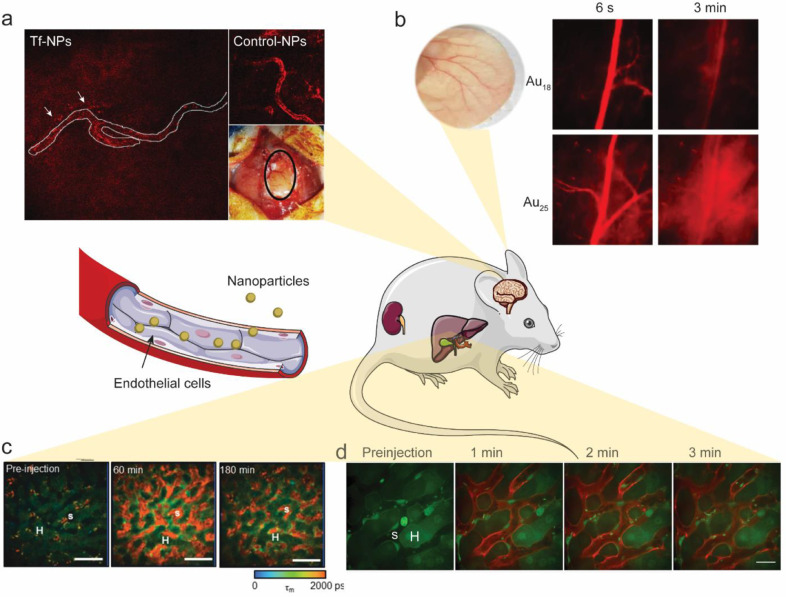
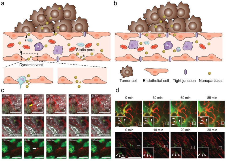
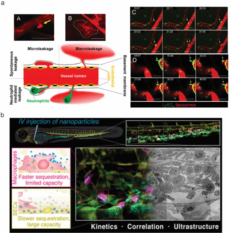
Nanomedicine has proven promising in preclinical studies. However, only few formulations have been successfully translated to clinical use. A thorough understanding of how nanoparticles interact with cells in vivo is essential to accelerate the clinical translation of nanomedicine. Intravital imaging is a crucial tool to reveal the mechanisms of nanoparticle transport in vivo, allowing for the development of new strategies for nanomaterial design. Here, we first review the most recent progress in using intravital imaging to answer fundamental questions about nanoparticle delivery in vivo. We then elaborate on how nanoparticles interact with different cell types and how such interactions determine the fate of nanoparticles in vivo. Lastly, we discuss ways in which the use of intravital imaging can be expanded in the future to facilitate the clinical translation of nanomedicine.

Keywords: Endothelial cells; Enhanced permeability and retention (EPR); Intravital microscopy (IVM); Macrophages; Neutrophils.


Conflict of interest statement

Declaration of Competing Interest The authors declare that there are no conflicts of interest.

Figures

Fig 1
Fig. 1
Intravital imaging sheds light on nanoparticle-endothelial cell interactions. a) Transferrin-functionalized nanoparticles (Tf-NPs: transferrin-PEG2K-Cy5.5 liposomes) cross the intact BBB, while control-NPs (Hemagglutinin-PEG2K-Cy5.5 liposomes) do not. Modified from Lam et al. with permission; b) Non-invasive bright-field and fluorescence images of mouse ear blood vessels 6 s and 3 min after i.v. injection of Au18 or Au25 clusters, showing that Au25 crossed the endothelium more rapidly than Au18. Adapted from Du et al. with permission; c) FLIM images of representative rat liver before, 60 min after, and 180 min after bolus injection of quantum dots (QD) with an emission channel of 515–620 nm. H represents the hepatocytes, while S represents the hepatic sinusoids. Adapted from Liang et al. with permission; d) Intravital imaging of α-melittin-NPs in the liver showed that NPs quickly target LSECs. α-melittin-NPs were labeled with DiR-BOA (red), a lipid-anchored near-infrared fluorophore. Actb-EGFP mice were used to visualize the structure of the liver. Adapted from Yu et al. with permission. (For interpretation of the references to color in this figure legend, the reader is referred to the web version of this article.)
Fig 2
Fig. 2
New insights on EPR using IVM. a) NP transport through gaps between adjacent endothelial cells in dynamic vascular bursts; b) NP transport across the endothelial cell layer via transcytosis. c) Representative eruption (white arrow) occurring near a Hoechst-stained (white) leukocyte cell (yellow arrow) (top) and an eruption occurring without cells nearby(middle), respectively. 70 nm nanoparticles (red) and a BxPC3-GFP dorsal skinfold model (green) were used. Eruption of Doxil particles (red) using a GFP dorsal skinfold model (green) (bottom). Scale bars, 100 μm. Adapted from Matsumoto et al. with permission; d) Intravital imaging shows colocalization of nanoparticles with endothelial cells to form hotspots along the vessel lining (red, stained with GSL1-Cy3). Arrows indicate hotspots. These vessels belong to MMTVPyMT (top) and 4T1 (bottom) tumour models. The 50 nm AuNPs (green) were conjugated with Alexa Fluor 647 to obtain the fluorescent signal. Scale bars, 200 μm; insets, 20 μm. Adapted from Sindhwani et al. with permission. (For interpretation of the references to color in this figure legend, the reader is referred to the web version of this article.)
Fig 3
Fig. 3
The collaborative and competitive relationship between NPs and cells. a) IVM reveals two different liposome extravasation patterns. A-B. Representative images of liposome extravasation in 4T1 orthotopic tumors through microleakage (A, arrow) and macroleakage (B, dashed line) after i.v. injection. Time-lapse imaging of neutrophil extravasation followed by microleakage (C, arrow) and macroleakage development (D,arrow); red, liposomes; green, Ly6G-positive cells; scale bar, 50 μm. Yellow arrow in C points to an extravasated neutrophil crawling in the perivascular area. Adapted from Naumenko et al. with permission; b) Faster nanoparticle sequestration by macrophages with limited capacity and slower nanoparticle sequestration via SECs with large capacity, visualized in vivo in real-time and at ultrastructural resolution. Adapted from Hayashi et al. with permission. (For interpretation of the references to color in this figure legend, the reader is referred to the web version of this article.)

References

    1. Anselmo A.C., Mitragotri S. Nanoparticles in the clinic: an update. Bioeng Transl Med. 2019;4(3):e10143. - PMC - PubMed
    1. Mitragotri S., Anderson D.G., Chen X., Chow E.K., Ho D., Kabanov A.V. Accelerating the translation of nanomaterials in biomedicine. ACS Nano. 2015;9(7):6644–6654. - PMC - PubMed
    1. Wilhelm S., Tavares A.J., Dai Q., Ohta S., Audet J., Dvorak H.F. Analysis of nanoparticle delivery to tumours. Nat Rev Mater. 2016;1(5):16014.
    1. Miller M.J., Wei S.H., Parker I., Cahalan M.D. Two-photon imaging of lymphocyte motility and antigen response in intact lymph node. Science. 2002;296(5574):1869–1873. - PubMed
    1. Stoll S., Delon J., Brotz T.M., Germain R.N. Dynamic imaging of T cell-dendritic cell interactions in lymph nodes. Science. 2002;296(5574):1873–1876. - PubMed