Review
. 2020 Aug 25;21(17):6109.
doi: 10.3390/ijms21176109.

Lamin A/C and the Immune System: One Intermediate Filament, Many Faces

Affiliations
Review

Lamin A/C and the Immune System: One Intermediate Filament, Many Faces

Angela Saez et al. Int J Mol Sci. .

Abstract

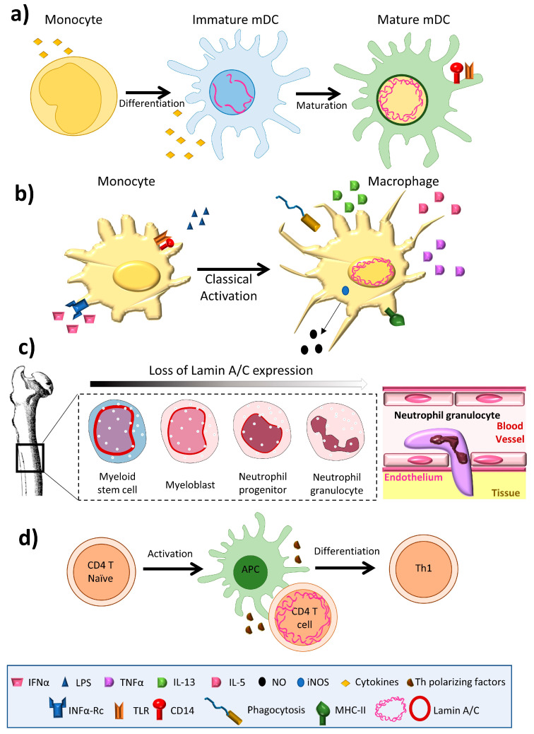
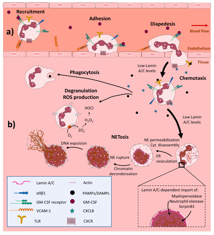
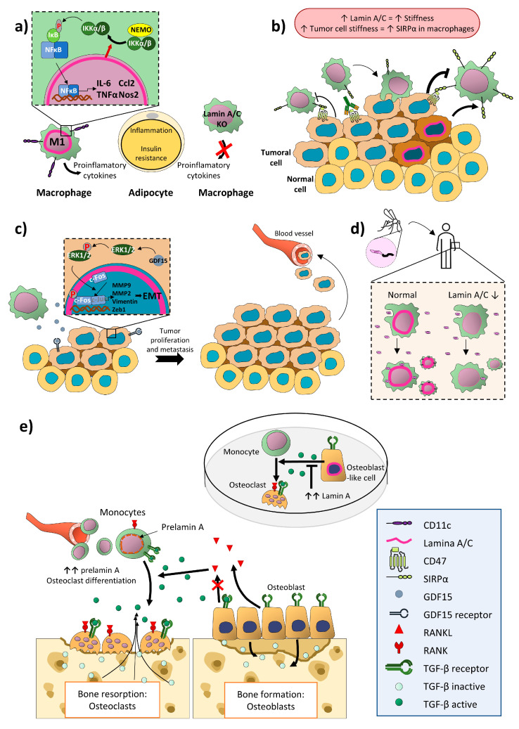
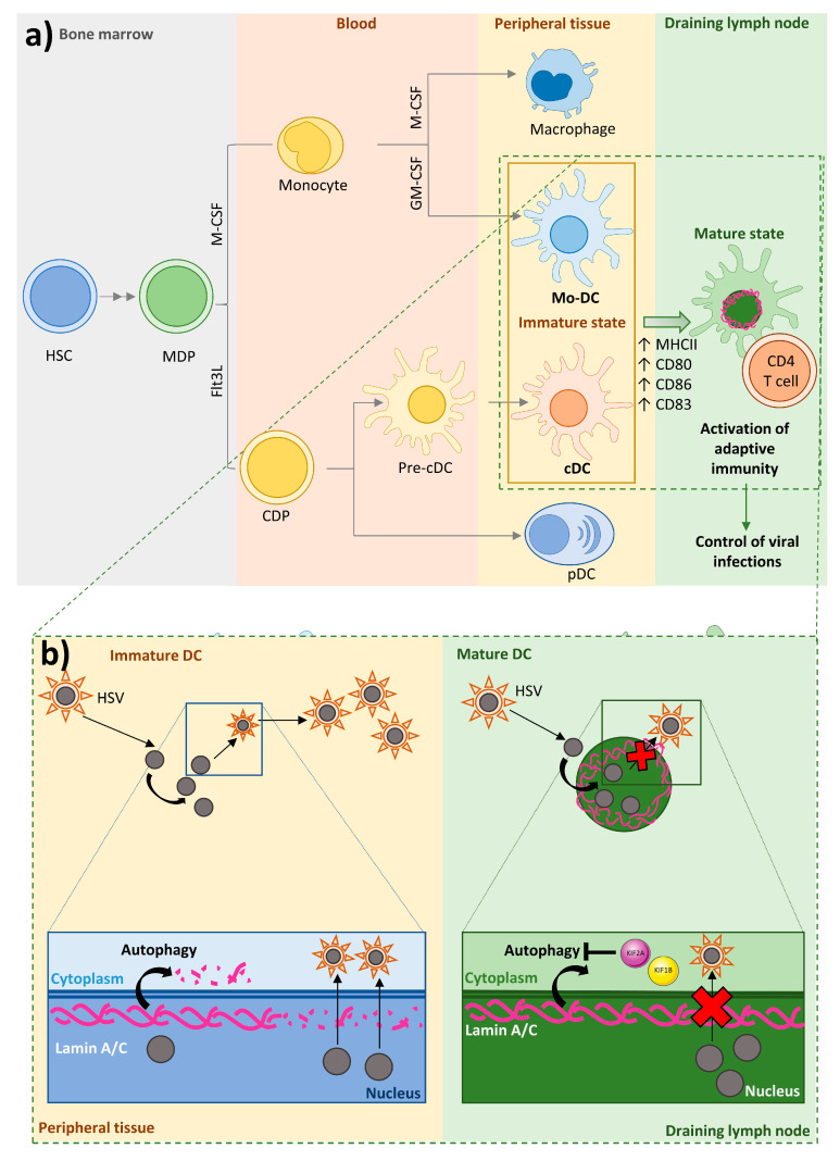
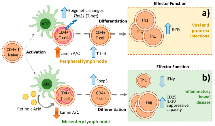
Nuclear envelope lamin A/C proteins are a major component of the mammalian nuclear lamina, a dense fibrous protein meshwork located in the nuclear interior. Lamin A/C proteins regulate nuclear mechanics and structure and control cellular signaling, gene transcription, epigenetic regulation, cell cycle progression, cell differentiation, and cell migration. The immune system is composed of the innate and adaptive branches. Innate immunity is mediated by myeloid cells such as neutrophils, macrophages, and dendritic cells. These cells produce a rapid and nonspecific response through phagocytosis, cytokine production, and complement activation, as well as activating adaptive immunity. Specific adaptive immunity is activated by antigen presentation by antigen presenting cells (APCs) and the cytokine microenvironment, and is mainly mediated by the cellular functions of T cells and the production of antibodies by B cells. Unlike most cell types, immune cells regulate their lamin A/C protein expression relatively rapidly to exert their functions, with expression increasing in macrophages, reducing in neutrophils, and increasing transiently in T cells. In this review, we discuss and summarize studies that have addressed the role played by lamin A/C in the functions of innate and adaptive immune cells in the context of human inflammatory and autoimmune diseases, pathogen infections, and cancer.

Keywords: Leishmania; T cell; cancer; dendritic cell (DC); inflammatory bowel disease; lamin A/C; macrophage; neutrophil; viral infection.


Conflict of interest statement

The authors declare no conflict of interest.

Figures

Figure 1
Figure 1
Lamin A/C levels are finely regulated in immune cells. (a) Dendritic cells have an intermediate lamin A/C content, between that of neutrophils and macrophages, which is associated with intermediate viability and migration. (b) Macrophages increase lamin A/C content upon differentiation and activation. (c) During granulopoiesis, neutrophil precursors change their round nuclear shape for a characteristic lobed nucleus, a process linked to almost complete loss of lamin A/C expression and augmented expression of the lamin B receptor (LBR). Neutrophil loss of lamin A/C enables them to pass through narrow spaces. (d) T cells show a transient peak in lamin A/C expression upon recognition of an antigen presented by an antigen presenting cell.
Figure 2
Figure 2
Low lamin A/C expression in neutrophils enables them to pass through narrow spaces during transendothelial migration. Upon infection or tissue damage, neutrophils are rapidly recruited from the circulation to tissues in response to chemoattractants such as CXCL8. The transmigration of neutrophils across the endothelium is mediated by interaction between integrin α9β1 expressed on neutrophils and VCAM-1 expressed on the activated endothelium. Stimulation of neutrophils by GM-CSF during recruitment augments their lifespan. Neutrophils recognize PAMPs or DAMPs through the activation of TLRs and contribute to the resolution of infection through phagocytosis, the release of granules and ROS, and the formation of neutrophil extracellular traps (NETs) NETosis. Neutrophils engage several cellular mechanisms during the generation of NETs (NETosis): disassembly of the cytoskeleton, endoplasmic reticulum vesiculation, chromatin decondensation, plasma membrane and nuclear envelope permeabilization, nuclear lamin meshwork and nuclear envelope rupture to release DNA to the cytoplasm, and plasma membrane rupture and expulsion of DNA to the extracellular environment. (a) The low content of lamin A/C in neutrophils allows them to distort their nuclei and pass through narrow spaces during transendothelial migration. (b) During NETosis, low lamin A/C content facilitates nuclear envelope rupture. This event also involves nuclear transport of cytoplasmic granule proteins, and low lamin A/C might be also facilitate in this process by easing the control of transport across nuclear pore complexes. GM-CSF, granulocyte-macrophage colony-stimulating factor; VCAM-1, vascular cell adhesion molecule 1; TLR, toll-like receptor; PAMPs, pathogen-associated molecular patterns; DAMPs, damage-associated molecular patterns; CXCL8, C-X-C motif chemokine ligand 8; CXCR, CXC chemokine receptor; ER, endoplasmic reticulum; NE, nuclear envelope; Cyt., Cytoskeleton.
Figure 3
Figure 3
Lamin A/C plays important roles in monocytes and macrophages in pathogen responses, cancer, and obesity. (a) Lamin A/C in M1 CD11c+ macrophages promotes obesity-induced insulin resistance and reduces adipose tissue inflammation by activating IKK, promoting NF-κB nuclear translocation, and increasing the expression of proinflammatory genes. (b) Expression of CD47 on tumor cells inhibits the action of macrophages by interacting with SIRPα. Lamin A/C content positively correlates with cell stiffness, and stiffness of solid tumors increases macrophage expression of SIRPα, reducing their phagocytic phenotype. The lamin A/C content of tumor macrophages may serve as an index of SIRPα content, potentially providing a means of predicting the efficiency of CD47- SIRPα checkpoint-directed immunotherapies in some cancer types. (c) Tumor macrophages produce GDF15, which in colorectal cancer cells releases c-Fos from inhibition by lamin A/C via ERK1/2 phosphorylation, resulting in transcription of AP1-regulated genes and subsequent tumor cell proliferation and metastasis. (d) Macrophages are important for resolving infection by Leishmania braziliensis, but can also be infected and serve as a niche for parasite replication. Lower lamin A/C content in macrophages diminishes the prevalence of L. braziliensis in these cells. (e) Bone mass remodeling depends on osteoblasts and osteoclasts. TGFβ stimulates monocytes to form osteoclasts, which mediate bone resorption, and indirectly limits further osteoclast formation by downregulating the osteoclast differentiation factor RANKL in osteoblasts. TGFβ thus regulates bone resorption by first promoting osteoclast differentiation from monocytes and then limiting the extent and duration of resorption. Increased accumulation of the lamin A precursor prelamin A in monocytes induces differentiation towards osteoclasts. In the osteoblast-like cell line U2-OS, Lamin A negatively modulates TGFβ 2 release, which may affect monocyte-to-osteoclast differentiation. T arrow, inhibition; red cross, blockade; up arrow, increase; down arrow, decrease.
Figure 4
Figure 4
DC ontogeny and the importance of DC-expressed lamin A/C in the response to HSV infection. (a) A common progenitor, the macrophage/dendritic cell progenitor (MDP), gives rise to macrophages, monocyte derived-DCs, conventional DCs, and plasmacytoid DCs. (b) DCs play an important role in the initiation of immune responses against pathogens through their function as professional APCs and their secretion of cytokines. Depending on their state of maturation, DCs are located in peripheral tissues (immature DCs) or in draining lymph nodes (mature DCs), to where they migrate after encountering and taking up antigens. In draining lymph nodes, DCs stimulate adaptive immune responses that help to control and destroy viral infection. Some viruses infect DCs, promoting the production, release, and spreading of viral particles in host cells and the host organism. In the case of herpex simplex virus (HSV), nuclear egress of progeny capsids, the formation of new infectious particles, and the viral release and spread depend on the nuclear lamina permeabilization and lamin A/C degradation by autophagy. Unlike immature DCs, mature DCs have an elevated protein content of the mTOR activators KIF1B and KIF2A, leading to inhibition of autophagy-dependent lamin A/C degradation after HSV infection and thus limiting viral nuclear egress, viral particle formation, and virus release and spread.
Figure 5
Figure 5
Lamin A/C in CD4+ T cells respectively promotes and inhibits the differentiation and function of Th1 cells and Tregs. Upon antigen recognition, CD4+ T cells activate, proliferate, and differentiate into specialized effector T helper (Th) cells. (a) In the context of viral or protozoan infection, lamin A/C expression increases CD4+ T cell Th1 differentiation by epigenetically regulating the mRNA expression of Tbx21 (T-bet). (b) In inflammatory bowel disease, the absence of lamin A/C in CD4 T cells enhances FOXP3 expression, which is reflected in increased Treg differentiation and function, important in resolving the impaired Th1 response. Vertical arrows indicate changes in expression or function.The efficacy of anti-tumor drugs depend on their capacity to induce apoptosis since apoptosis evasion is a redundant characteristic of cancer cells [223]. During apoptosis, the nuclear envelope is permeabilized by rupture of nuclear pore complex and nuclear lamina proteins, including lamin A/C [224]. Classical Hodgkin’s lymphoma is a kind of B cell lymphoma contained of Hodgkin and Reed-Sternberg cells with a unique or multiple nucleus, respectively [17]. Hodgkin and Reed-Sternberg cells show lamin A/C expression [33] and impaired lamin A/C structure, which is dissimilar from unstimulated common lymphocytes [17]. It has been described a caspase-dependent and independent [224] cleavage of lamin A/C, which dismantles the nuclear lamina, and promotes apoptosis in B cell lymphomas [225,226].

References

    1. Broers J.L., Ramaekers F.C., Bonne G., Yaou R.B., Hutchison C.J. Nuclear lamins: Laminopathies and their role in premature ageing. Physiol. Rev. 2006;86:967–1008. doi: 10.1152/physrev.00047.2005. - DOI - PubMed
    1. Gonzalez J.M., Pla D., Perez-Sala D., Andres V. A-type lamins and Hutchinson-Gilford progeria syndrome: Pathogenesis and therapy. Front. Biosci. (Schol. Ed.) 2011;3:1133–1146. doi: 10.2741/216. - DOI - PubMed
    1. Dechat T., Adam S.A., Taimen P., Shimi T., Goldman R.D. Nuclear lamins. Cold Spring Harb. Perspect. Biol. 2010;2:a000547. doi: 10.1101/cshperspect.a000547. - DOI - PMC - PubMed
    1. Moir R.D., Yoon M., Khuon S., Goldman R.D. Nuclear lamins A and B1: Different pathways of assembly during nuclear envelope formation in living cells. J. Cell Biol. 2000;151:1155–1168. doi: 10.1083/jcb.151.6.1155. - DOI - PMC - PubMed
    1. Andres V., Gonzalez J.M. Role of A-type lamins in signaling, transcription, and chromatin organization. J. Cell Biol. 2009;187:945–957. doi: 10.1083/jcb.200904124. - DOI - PMC - PubMed

LinkOut - more resources