Review
. 2020 Aug 25;21(17):6129.
doi: 10.3390/ijms21176129.

Elucidation of Melanogenesis Cascade for Identifying Pathophysiology and Therapeutic Approach of Pigmentary Disorders and Melanoma

Affiliations
Review

Elucidation of Melanogenesis Cascade for Identifying Pathophysiology and Therapeutic Approach of Pigmentary Disorders and Melanoma

Tokimasa Hida et al. Int J Mol Sci. .

Abstract

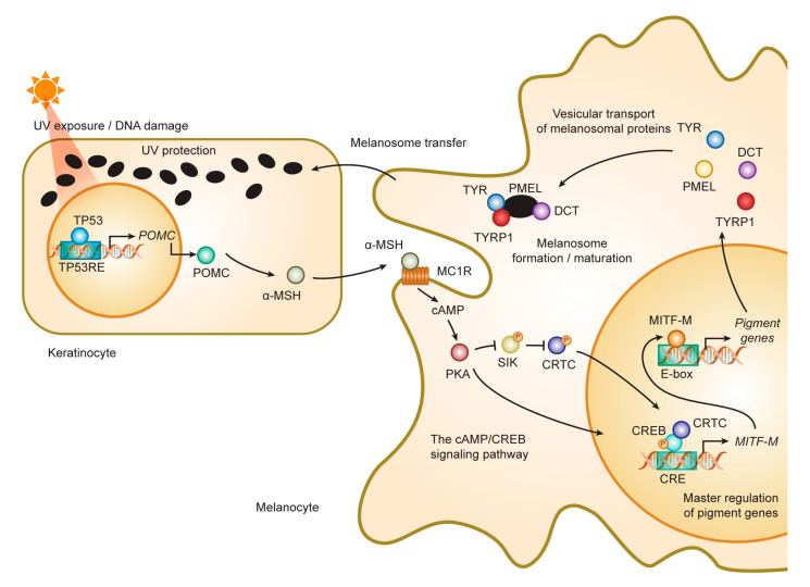
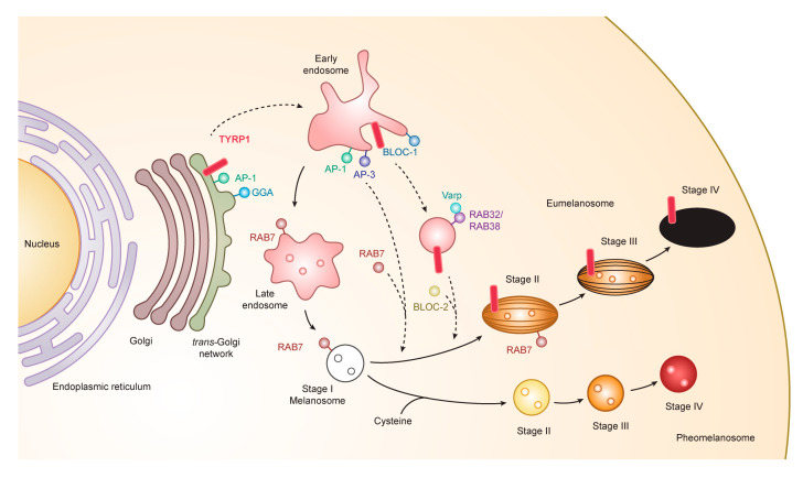
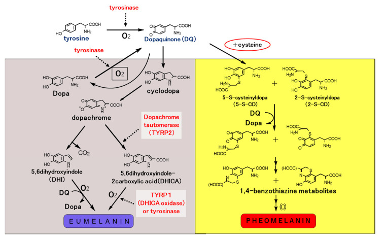
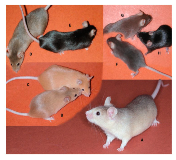
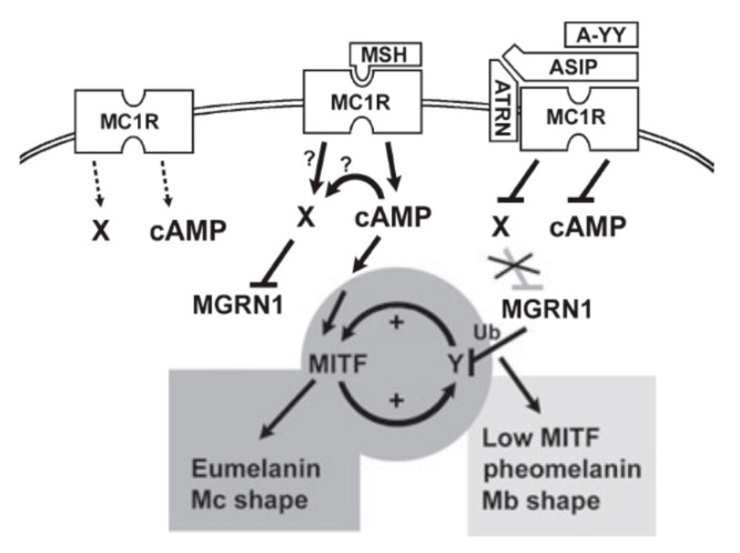
Melanogenesis is the biological and biochemical process of melanin and melanosome biosynthesis. Melanin is formed by enzymic reactions of tyrosinase family proteins that convert tyrosine to form brown-black eumelanin and yellow-red pheomelanin within melanosomal compartments in melanocytes, following the cascades of events interacting with a series of autocrine and paracrine signals. Fully melanized melanosomes are delivered to keratinocytes of the skin and hair. The symbiotic relation of a melanocyte and an associated pool of keratinocytes is called epidermal melanin unit (EMU). Microphthalmia-associated transcription factor (MITF) plays a vital role in melanocyte development and differentiation. MITF regulates expression of numerous pigmentation genes for promoting melanocyte differentiation, as well as fundamental genes for maintaining cell homeostasis. Diseases involving alterations of EMU show various forms of pigmentation phenotypes. This review introduces four major topics of melanogenesis cascade that include (1) melanocyte development and differentiation, (2) melanogenesis and intracellular trafficking for melanosome biosynthesis, (3) melanin pigmentation and pigment-type switching, and (4) development of a novel therapeutic approach for malignant melanoma by elucidation of melanogenesis cascade.

Keywords: eumelanin; hypomelanosis; melanogenesis; melanoma; melanosome; pheomelanin; pigment-type switching; tyrosinase; tyrosinase related protein (TYRP); vesicular transport.


Conflict of interest statement

The authors declare that they have no conflict of interest. The funders had no role in the design of the study; in the collection, analyses, or interpretation of data; in the writing of the manuscript; or in the decision to publish the results.

Figures

Figure 1
Figure 1
Role of MITF in melanogenesis cascade in human skin. The cascade of melanogenesis consists of the interaction between a melanocyte and an associated pool of keratinocytes, called epidermal melanin unit. MITF plays a master role in the cascade process for the activation/differentiation of the melanocyte followed by the induction of melanogenesis cascade such as during UV-induced tanning reaction. UV damages DNA in keratinocytes. DNA damage activates TP53 that initiates the transcription of POMC. POMC is cleaved into peptides including α-MSH. α-MSH secreted from keratinocytes binds MC1R and increases cAMP in melanocytes. PKA activated by cAMP activates CREB transcriptional activity in two ways: (i) Phosphorylate CREB; (ii) inhibiting SIK, a negative regulator of CREB co-activator CRTCs, by phosphorylating SIK. CREB activates the transcription of MITF-M. MITF-M activates the transcription of pigment genes including TYR, TYRP1, DCT, and PMEL. Melanosomal proteins are delivered to melanosomes. Matured melanosomes are transferred from melanocytes to keratinocytes and accumulate on the nucleus to block UV light. Abbreviations: α-MSH, alpha-melanocyte-stimulating hormone; cAMP, cyclic adenosine monophosphate; CRE, cAMP response element; CREB, CRE-binding protein; CRTC, CREB-regulated transcriptional coactivator; MC1R, melanocortin 1 receptor; MITF, microphthalmia-associated transcription factor; SIK, salt-inducible kinase; PKA, protein kinase A; POMC, proopiomelanocortin; TYR, tyrosinase; TYRP1, tyrosinase-related protein 1.
Figure 2
Figure 2
Melanogenesis and intracellular trafficking for melanosome biosynthesis. MITF turns on melanogenesis by activating transcription of pigment genes such as TYR, TYRP1, DCT, and PMEL which are transported from the trans-Golgi network and incorporated into early/late endosomal compartment to form melanosomes. Melanosomes have four maturation stages characterized by unique shapes and amounts of melanin pigments, i.e., eumelanin and pheomelanin. In the case of TYRP1, AP-1 and GGA play key roles in the sorting process of export from trans-Golgi network via early/late endosomes to stage I melanosomes by two other mechanisms: (i) BLOC-1/RAB32/RAB38/Varp/BLOC-2; (ii) AP-1 or AP-3. RAB7 is required for the stage I melanosome formation and the TYRP1 sorting from early/late endosomes to stage I melanosomes. Abbreviations: AP, adaptor protein; BLOC, biogenesis of lysosome-related organelles complex; GGA, Golgi-localized γ-ear-containing ADP-ribosylation factor -binding protein.
Figure 3
Figure 3
Biosynthetic pathway of eumelanin and pheomelanin. Eumelanin and pheomelanin begin with the common pathway by converting tyrosine to Dopa and subsequently Dopa to dopaquinone. Tyrosinase interacts with two tyrosinase related proteins, TYRP1 and TYRP2 or dopachrome tautomerase (DCT) to form eumelanin pigments. On the other hand, cysteine is incorporated into dopaquinone to form cysteinyldopa which is then converted to benzothiazine metabolites to form pheomelanin pigments. Dopaquinone obtained by tyrosinase oxidation is a highly reactive intermediate, and in the absence of thiol compounds, it undergoes intramolecular cyclization, leading eventually to the production of eumelanin. However, the intervention of thiols, such as cysteine, with this process gives rise to the thiol adducts. Further oxidation of these cysteinyldopa isomers leads to the production of pheomelanin. In reality, most of the melanin pigments present in the hair and skin, and in melanomas may not be homopolymers of a single monomer unit, but rather they are complex heteropolymers made up of both eumelanin and pheomelanin building blocks.
Figure 4
Figure 4
Cascade of eumelanogenesis and pheomelanogenesis. Mature stage IV melanosomes reveal a unique shape. Eumelanosomes become ellipsoidal with specific lattice-like internal structure that is formed by structural matrix protein, PMEL. Pheomelanosomes take oval forms with many internal structures. Both eumelanosomes and pheomelanosomes possess a significant number of microvesicles, which were originally reported as “vesiculoglobular bodies” deriving from the carriers of vesicular transport [41].
Figure 5
Figure 5
Mouse color mutations. All mice are of strain C57BL/6J except (A). On the C57BL/6J background, the Ay/a mouse is clear yellow (C), whereas on the JU/CtLm background it is paler clear yellow (A). (B) Mc1re/Mc1re (recessive yellow). (D) Ay/a, Atrnmg/Atrnmg (mahogany). (E) a/a, Atrnmg/Atrnmg. (F) Dctslt/Dctslt, slaty. (G) Tyrp1b/Tyrp1b, brown. (H) C57BL/6J (a/a, nonagouti), control mouse. Reproduced from reference [72], courtesy of Bennett, D.C. and Lamoreux, M.L.
Figure 6
Figure 6
Model of pigment-type switching controlled by dual signaling from MC1R. ASIP, agouti-signaling protein (agouti protein); A-YY (ASIP-YY), a synthetic C-terminal fragment of agouti protein; Mc, melanocyte; Mb, melanoblast; Ub, ubiquitinylation. Melanocortin-1 receptor (MC1R) is shown with some basal activity (left). MC1R can activate both cAMP and another signaling pathway X. Activation of X may be cAMP-dependent or -independent. Agouti protein can antagonize cAMP signaling through its C-terminus (or as ASIP-YY) and can independently antagonize X signaling through its N-terminus and through ATRN and MGRN1, resulting in inhibition of eumelanogenesis and enhancement of pheomelanogenesis. To explain the activation of eumelanogenesis by cAMP, yet persistence of eumelanogenesis when the cAMP signal is withdrawn, a positive feedback loop was suggested. MGRN1 may break the loop by ubiquitinating and destabilizing one component Y. This would lead to a less differentiated state with low MITF activity, permissive for pheomelanogenesis. Reproduced from reference [78].
Figure 7
Figure 7
Depigmenting effect of N-acetyl-4-S-cysteaminylphenol (NAcCAP). Marked depigmentation of facial pigmentation in a melasma patient after daily topical application of NAcCAP ointment [96].
Figure 8
Figure 8
Synthesis of N-propionyl-4-S-cysteaminylphenol/magnetite nanoparticles. Several forms of N-propionyl-4-S-cysteaminylphenol (NPrCAP)/magnetite (M) nanoparticles conjugates have been synthesized. Form 1: NPrCAP is conjugated on the surface of neutral magnetic liposome particles (NML). Form 2: NPrCAP is embedded in cationic magnetic liposome particles (CML). Form 3: NPrCAP is directly conjugated with magnetite. Form 4: NPrCAP is conjugated with magnetite by polyethylene glycol (PEG).
Figure 9
Figure 9
Aggregation of NPrCAP/M in melanosomes of B16 melanoma cells. The electron microscopic examination shows selective delivery of NPrCAP/M into melanosomes 14 days after intraperitoneal administration. M, magnetite.
Figure 10
Figure 10
Protocol and mechanism of CTI therapy for melanoma patients in the advanced stage. STEP 1: NPrCAP/PEG/M is injected directly into the target lesion (a skin metastasis on the body surface), followed by AMF irradiation to induce intratumoral hyperthermia at 43 °C for 30 min. STEP 2: Non-apoptotic cell death is induced in the majority of melanoma cells by hyperthermia. STEP 2’: Melanoma cells surviving from the hyperthermia treatment undergo apoptotic cell death through the chemotherapeutic effect of NPrCAP. STEP 3: HSPs and antigenic peptides released from dead cells are taken up by antigen-presenting cells. STEP 4: Anti-tumor immunity acquired through hyperthermia or unknown effects of NPrCAP induces migration of CD8+ T cells to kill other metastases.

References

    1. Jimbow K., Prota G., Quevedo W.C., Fitzpatrick T.B. Biology of Melanocytes. In: Freedberg I.M., Eisen A.Z., Wolff E., Austen K.F., Goldsmith L.A., Katz S.I., Fitzpatrick T.B., editors. Fitzpatrick’s Dermatology in General Medicine. 5th ed. McGraw-Hill; New York, NY, USA: 1998. pp. 192–220.
    1. Kawakami A., Fisher D.E. The master role of microphthalmia-associated transcription factor in melanocyte and melanoma biology. Lab. Investig. 2017;97:649–656. doi: 10.1038/labinvest.2017.9. - DOI - PubMed
    1. Toyofuku K., Wada I., Hirosaki K., Park J.S., Hori Y., Jimbow K. Promotion of tyrosinase folding in Cos 7 cells by calnexin. J. Biochem. 1999;125:82–89. doi: 10.1093/oxfordjournals.jbchem.a022272. - DOI - PubMed
    1. Jimbow K., Park J.S., Kato F., Hirosaki K., Toyofuku K., Hua C., Yamashita T. Assembly, target-signaling and intracellular transport of tyrosinase gene family protein in the initial stage of melanosome biogenesis. Pigment Cell Res. 2000;13:222–229. doi: 10.1034/j.1600-0749.2000.130403.x. - DOI - PubMed
    1. Kamada A., Nagaya H., Tamura T., Kinjo M., Jin H.Y., Yamashita T., Jimbow K., Kanoh H., Wada I. Regulation of immature protein dynamics in the endoplasmic reticulum. J. Biol. Chem. 2004;279:21533–21542. doi: 10.1074/jbc.M401403200. - DOI - PubMed

MeSH terms

Substances