Comparative Study
. 2020 Aug 25;21(17):6132.
doi: 10.3390/ijms21176132.

Expansion of Human Limbal Epithelial Stem/Progenitor Cells Using Different Human Sera: A Multivariate Statistical Analysis

Affiliations
Comparative Study

Expansion of Human Limbal Epithelial Stem/Progenitor Cells Using Different Human Sera: A Multivariate Statistical Analysis

Raquel Hernáez-Moya et al. Int J Mol Sci. .

Abstract

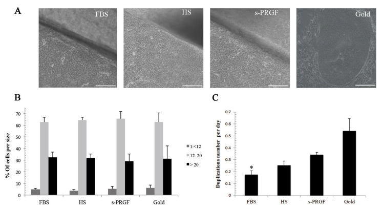
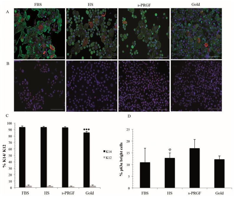
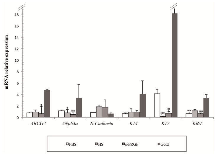
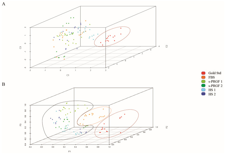
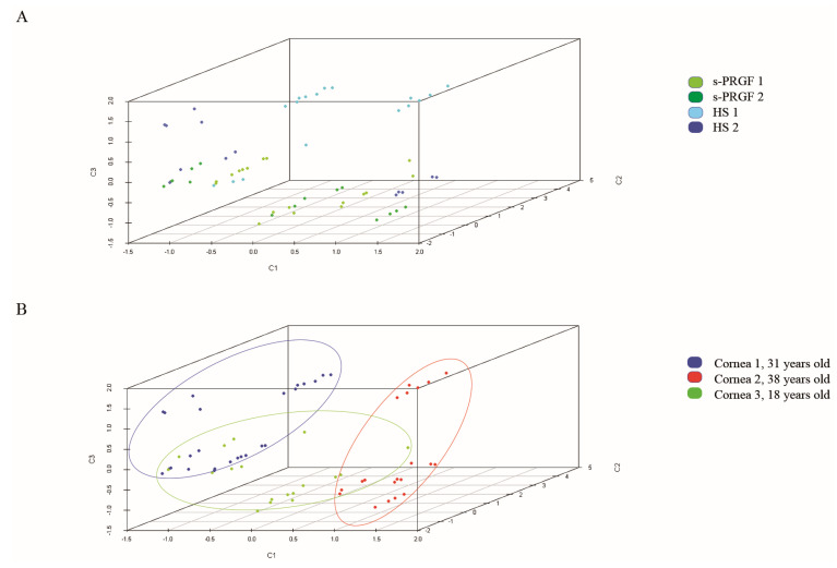
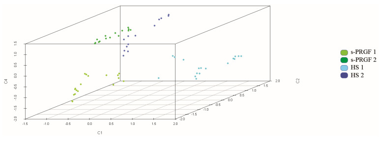
Transplantation of human cultured limbal epithelial stem/progenitor cells (LESCs) has demonstrated to restore the integrity and functionality of the corneal surface in about 76% of patients with limbal stem cell deficiency. However, there are different protocols for the expansion of LESCs, and many of them use xenogeneic products, being a risk for the patients' health. We compared the culture of limbal explants on the denuded amniotic membrane in the culture medium-supplemental hormone epithelial medium (SHEM)-supplemented with FBS or two differently produced human sera. Cell morphology, cell size, cell growth rate, and the expression level of differentiation and putative stem cell markers were examined. Several bioactive molecules were quantified in the human sera. In a novel approach, we performed a multivariate statistical analysis of data to investigate the culture factors, such as differently expressed molecules of human sera that specifically influence the cell phenotype. Our results showed that limbal cells cultured with human sera grew faster and contained similar amounts of small-sized cells, higher expression of the protein p63α, and lower of cytokeratin K12 than FBS cultures, thus, maintaining the stem/progenitor phenotype of LESCs. Furthermore, the multivariate analysis provided much data to better understand the obtaining of different cell phenotypes as a consequence of the use of different culture methodologies or different culture components.

Keywords: cell therapy; explants culture; human amniotic membrane (HAM); human serum (HS); limbal epithelial stem/progenitor cells (LESC); limbal stem cell deficiency (LSCD); multivariate statistical analysis; partial least squares discriminant analysis (PLS-DA); principal component analysis (PCA) combined with clusters analysis; serum derived from plasma rich in growth factors (s-PRGF); xenogeneic-free cell expansion.


Conflict of interest statement

No sponsor or funding organization had any role in the design or conduct of this research. None of the authors had any conflict of interest.

Figures

Figure 1
Figure 1
LESCs cultured following the gold standard or explants culture methodologies with FBS or human HS and s-PRGF sera: (A) Phenotype of cell cultures; (B) Percentage of cells corresponding to each cell size; (C) Cell growth rate represented as the number of cell duplications per day. * Indicates significant differences between the gold standard culture and the indicated treatment. (* p < 0.05). Scale bar in phase-contrast microscopy images, 100 micrometers. LESCs: limbal epithelial stem/progenitor cells; FBS: fetal bovine serum; HS: human serum; s-PRGF: serum derived from plasma rich in growth factors.
Figure 2
Figure 2
Protein expression profiles of several cell markers in LESCs obtained from explants and gold standard cultures treated with FBS or two different human sera: (A) Double immunostaining for K14 (green) and K12 (red) cytokeratins; (B) Immunostaining for p63α; (C) Percentage of K14 and K12 positive cells; (D) Percentage of p63α bright cells. *** Indicates significant differences between the gold standard culture and all the other treatments (p < 0.001), and φ between FBS and the indicated treatment (p < 0.05). Scale bar for fluorescence images, 100 micrometers.
Figure 3
Figure 3
Relative mRNA expression of several cell markers in LESCs obtained from explants and gold standard cultures treated with FBS or two different human sera. * Indicates significant differences between the gold standard culture and the indicated treatments, and φ between the FBS and the indicated treatments. (* p < 0.05; ** p < 0.01) (φ p < 0.05; φφ p < 0.01).
Figure 4
Figure 4
Representation of all samples in three components obtained by (A) Principal component analysis (PCA); (B) Partial least squares discriminant analysis (PLS-DA). Colors indicate treatments. Cultures following the gold standard culture methodology are separated from cultures following explants culture methodology.
Figure 5
Figure 5
Representation of samples treated with human sera in three components obtained by PCA analysis. Colors indicate (A) treatments; (B) group of cornea-HAM (human amniotic membrane). Samples were classified mainly by cornea-HAM and not by treatment.
Figure 6
Figure 6
Representation of samples treated with human sera in three components obtained by PCA analysis. The analysis also included as variables some molecules measured in sera (glial cell-derived neurotrophic factor (GDNF), substance P (SP), epidermal growth factor (EGF)). Colors indicate treatments. Samples were well classified by treatment.

Similar articles

Cited by

References

    1. Davanger M., Evensen A. Role of the pericorneal papillary structure in renewal of corneal epithelium. Nature. 1971;229:560–561. doi: 10.1038/229560a0. - DOI - PubMed
    1. Tseng S.C.G. Concept and application of limbal stem cells. Eye. 1989;3:141–157. doi: 10.1038/eye.1989.22. - DOI - PubMed
    1. Schlötzer-Schrehardt U., Kruse F.E. Identification and characterization of limbal stem cells. Exp. Eye Res. 2005;81:247–264. doi: 10.1016/j.exer.2005.02.016. - DOI - PubMed
    1. Seyed-Safi A.G., Daniels J.T. The limbus: Structure and function. Exp. Eye Res. 2020;197:108074. doi: 10.1016/j.exer.2020.108074. - DOI - PubMed
    1. Dua H.S., Shanmuganathan V.A., Powell-Richards A.O., Tighe P.J., Joseph A. Limbal epithelial crypts: A novel anatomical structure and a putative limbal stem cell niche. Br. J. Ophthalmol. 2005;89:529–532. doi: 10.1136/bjo.2004.049742. - DOI - PMC - PubMed

Publication types

MeSH terms