

Neurosarcoidosis presenting as CRVO combined CRAO: a biopsy-proven case report of a Chinese patient
- PMID: 32854651
- PMCID: PMC7457306
- DOI: 10.1186/s12886-020-01624-5
Neurosarcoidosis presenting as CRVO combined CRAO: a biopsy-proven case report of a Chinese patient
Abstract
Background: Neurosarcoidosis is a rare systemic disorder that can affect the eye and other organs, including the central nervous system. Neurosarcoidosis infiltrating the optic nerve presenting as central retinal vein occlusion combined with artery ischaemia has not been reported in the literature previously. We describe a Chinese patient presenting with acute monocular vision loss, in whom an optic nerve biopsy confirmed the diagnosis of neurosarcoidosis.
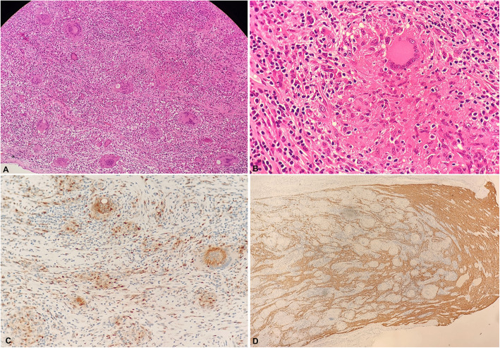
Case presentation: A 47-year-old woman complained of acute decreased vision in her left eye over the course of 1 month. She reported that her vision deteriorated quickly within first 3 days of consulting an ophthalmologist at a local hospital. She was diagnosed with central retinal vein occlusion after funduscopic examination and fundus fluorescein angiography, and the vision in her left eye further deteriorated to no light perception. An orbital magnetic resonance imaging showed an abnormal T1-weighted image of the optic nerve after contrast enhancement. She was referred to a neuro-ophthalmologist for further evaluation. After routine blood tests ruled out infectious and metastatic diseases, she was prescribed 500 mg/d methylprednisolone for 5 days, but her vision did not improve. As she could still not perceive light, an optic nerve biopsy was performed, and the histopathology revealed non-necrotising granuloma that was consistent with neurosarcoidosis.
Conclusions: Isolated optic nerve infiltration by neurosarcoidosis without the involvement of the central nervous system or other systemic organs is challenging to diagnose. Biopsy of the optic nerve sheath is crucial for the final diagnosis of neurosarcoidosis. Therefore, a comprehensive ophthalmologic and systemic examination and work-up for inflammation of the eye, chest, and central nervous system should be conducted for atypical cases.
Keywords: Neurosarcoidosis; Optic nerve biopsy; Optic neuropathy; Retinal vein occlusion.
Conflict of interest statement
The authors declare that they have no competing interests.
Figures


References
Publication types
MeSH terms
Supplementary concepts
Grants and funding
LinkOut - more resources
Full Text Sources
Medical
Research Materials
