. 2020 Aug 27;20(1):48.
doi: 10.1186/s12896-020-00641-y.

Synergistic interaction of hTGF-β3 with hBMP-6 promotes articular cartilage formation in chitosan scaffolds with hADSCs: implications for regenerative medicine

Affiliations

Synergistic interaction of hTGF-β3 with hBMP-6 promotes articular cartilage formation in chitosan scaffolds with hADSCs: implications for regenerative medicine

Yijiang Huang et al. BMC Biotechnol. .

Abstract

Background: Human TGF-β3 has been used in many studies to induce genes coding for typical cartilage matrix components and accelerate chondrogenic differentiation, making it the standard constituent in most cultivation media used for the assessment of chondrogenesis associated with various stem cell types on carrier matrices. However, in vivo data suggests that TGF-β3 and its other isoforms also induce endochondral and intramembranous osteogenesis in non-primate species to other mammals. Based on previously demonstrated improved articular cartilage induction by a using hTGF-β3 and hBMP-6 together on hADSC cultures and the interaction of TGF- β with matrix in vivo, the present study investigates the interaction of a chitosan scaffold as polyanionic polysaccharide with both growth factors. The study analyzes the difference between chondrogenic differentiation that leads to stable hyaline cartilage and the endochondral ossification route that ends in hypertrophy by extending the usual panel of investigated gene expression and stringent employment of quantitative PCR.

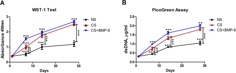
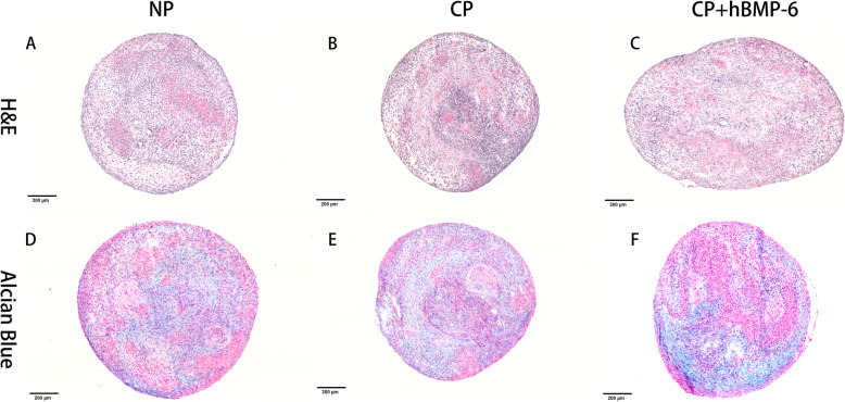
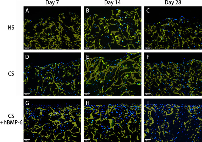
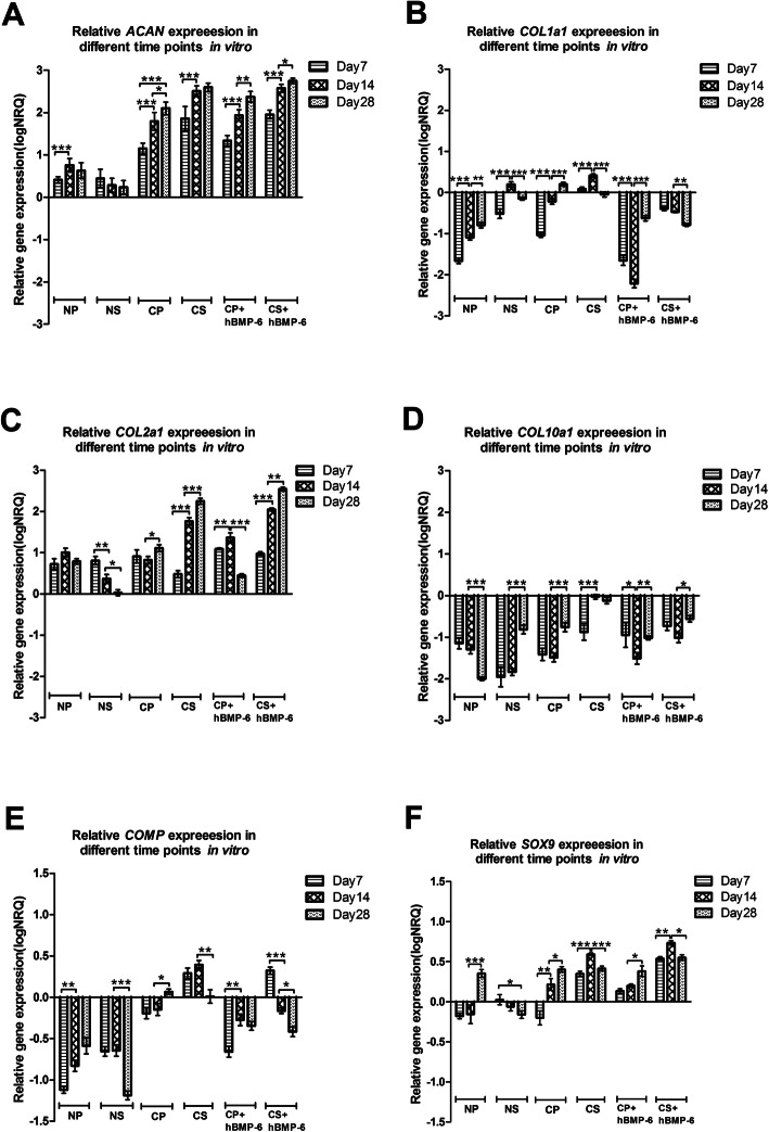
Results: By assessing the viability, proliferation, matrix formation and gene expression patterns it is shown that hTGF-β3 + hBMP-6 promotes improved hyaline articular cartilage formation in a chitosan scaffold in which ACAN with Col2A1 and not Col1A1 nor Col10A1 where highly expressed both at a transcriptional and translational level. Inversely, hTGF-β3 alone tended towards endochondral bone formation showing according protein and gene expression patterns.

Conclusion: These findings demonstrate that clinical therapies should consider using hTGF-β3 + hBMP-6 in articular cartilage regeneration therapies as the synergistic interaction of these morphogens seems to ensure and maintain proper hyaline articular cartilage matrix formation counteracting degeneration to fibrous tissue or ossification. These effects are produced by interaction of the growth factors with the polysaccharide matrix.

Keywords: Adipose-derived stem cell; Articular Chondrogenesis; Bone formation; Chitosan; Promotion; Synergism; Validation; hBMP-6; hTGF-β3.


Conflict of interest statement

The authors declare no conflict of interest.

Figures

Fig. 1
Fig. 1
Scaffold characteristics and cell distribution in culture with hADSCs at different time points. a Stereomicroscopic image of the dry scaffold, showing sponge-like uniform porous structure. b Scanning electron microscopy images of chitosan scaffolds before and (c) after incubation with medium (cell-free blank). Image in B was used for pore size determination. Images d-s demonstrate cellular growth on days 1 (d-g), 7 (h-k), 14 (l-o) and 28 (p-s), the first two images showing growth with hTGF-β3 only, the second with hTGF-β3 + hBMP-6. Grey-scale images (first and third column) are SEM images, fluorescent images (second and fourth column) are live/dead stain with green = Calcein AM for live and red = Ethidium bromide for dead cells. Collagenous fibrous matrix (black arrows) (h-k) For (a) magnification was set at 4x; SEM magnifications were set at 100x (b), 300x (c, d, h, j, l, p, r), 1.10Kx (f, n); Live/Dead magnifications were set at 10x (e, g, i, k, m, o, q, s)
Fig. 2
Fig. 2
a WST-1 (cell viability) and b PicoGreen (cell proliferation) assays for hADSCs on chitosan scaffolds cultured with normal (NS), standard chondrogenic (CS) or modified chondrogenic + hBMP-6 (CS + hBMP-6) medium. (***p < 0.001)
Fig. 3
Fig. 3
Alcian blue staining for GAG in chitosan scaffolds seeded with hADSCs cultured in either normal (NS) (a-c), standard chondrogenic (CS) (d-f) or modified chondrogenic + hBMP-6 medium (CS + hBMP-6) (g-i) after 7, 14 and 28 days. Magnification 40x (Bar scales: 200 μm)
Fig. 4
Fig. 4
H&E and Alcian blue staining of 3D hADSCs pellets cultured in either normal (a, d), standard chondrogenic (b, e) and modified chondrogenic + hBMP-6 medium (c, f) at day 28. H&E staining of the 3D hADSCs pellet cultures in normal medium (NP). Magnification 20x (Bar scales: 200 μm)
Fig. 5
Fig. 5
Histomorphometrical assessment of GAG (alcian blue) staining percentage between NS, CS and CS + hBMP-6 groups. (*p < 0.05, **p < 0.01, ***p < 0.001)
Fig. 6
Fig. 6
Immunofluorescence staining of aggrecan (green) at day 7, 14 and 28 in chitosan scaffolds with hADSCs cultured in normal (NS), standard chondrogenic (CS) or modified chondrogenic + hBMP-6 medium (CS + hBMP-6). The chitosan scaffolds fluoresced yellow, whereas living cell nuclei fluoresced blue. Magnification set a 10x
Fig. 7
Fig. 7
Immunofluorescence staining of collagen type II (green) at day 7, 14 and 28 in chitosan scaffolds with hADSCs cultured in normal (NS), standard chondrogenic (CS) or modified chondrogenic + hBMP-6 medium (CS + hBMP-6) . The chitosan scaffolds fluoresced yellow, whereas living cell nuclei fluoresced blue. Magnification set a 10x
Fig. 8
Fig. 8
Immunofluorescence staining of collagen type I (green) at day 7, 14 and 28 in chitosan scaffolds with hADSCs cultured in normal (NS), standard chondrogenic (CS) or modified chondrogenic + hBMP-6 medium (CS + hBMP-6). The chitosan scaffolds fluoresced yellow, whereas living cell nuclei fluoresced blue. Magnification set a 10x
Fig. 9
Fig. 9
Relative gene expression quantity of (a) ACAN, (b) COL1A1, (c) COL2A1, (d) COL10A1, (e) COMP and (f) SOX9 between all culture groups (N = normal medium; C = chondrogenic medium, P = 3D Pellet; S = chitosan scaffolds,). (*p < 0.05, **p < 0.01, ***p < 0.001). The baseline 0 represents untreated hADSCs in monolayer, which was the normalisation factor

Similar articles

Cited by

References

    1. Makris EA, Gomoll AH, Malizos KN, Hu JC, Athanasiou KA. Repair and tissue engineering techniques for articular cartilage. Nat Rev Rheumatol. 2015;11:21–34. doi: 10.1038/nrrheum.2014.157. - DOI - PMC - PubMed
    1. Hunter, W. On the structure and diseases of articulating cartilage. Phil Trans R Soc A 1743, 42B, 514–521.
    1. Marijnissen AC, Lafeber FP. Re: E. B. Hunziker. Articular cartilage repair: basic science and clinical progress. A review of the current status and prospects. Osteoarthritis and cartilage 2002; 10:432-63. Osteoarthr Cartil. 2003;11:300–301. doi: 10.1016/S1063-4584(02)00346-1. - DOI - PubMed
    1. Mandelbaum BR, Browne JE, Fu F, Micheli L, Mosely JB, Jr, Erggelet C, Minas T, Peterson L. Articular cartilage lesions of the knee. Am J Sports Med. 1998;26:853–861. doi: 10.1177/03635465980260062201. - DOI - PubMed
    1. Johnstone B, Alini M, Cucchiarini M, Dodge GR, Eglin D, Guilak F, Madry H, Mata A, Mauck RL, Semino CE, et al. Tissue engineering for articular cartilage repair--the state of the art. Eur Cell Mater. 2013;25:248–267. doi: 10.22203/eCM.v025a18. - DOI - PubMed

Publication types

MeSH terms

LinkOut - more resources