. 2020 Aug 27;18(1):117.
doi: 10.1186/s12951-020-00666-7.

MMP-13 enzyme and pH responsive theranostic nanoplatform for osteoarthritis

Affiliations

MMP-13 enzyme and pH responsive theranostic nanoplatform for osteoarthritis

Qiumei Lan et al. J Nanobiotechnology. .

Abstract

Stimulus-responsive therapy permits precise control of therapeutic effect only at lesion of interest, which determines it a promising method for diagnosis and imaging-guided precision therapy. The acid environment and overexpressed matrix metalloproteinases-13 (MMP-13) are typical markers in osteoarthritis (OA), which enables the development of stimulus-responsive drug delivery system with high specificity for OA. We herein demonstrate a nano-micelle based stimuli-responsive theranostic strategy with reporting and drug release controlled by acidic pH and MMP-13 for OA therapy. Such nanoplatform is incorporated with a motif specifically targeting on cartilage, a motif responsive to matrix metalloproteinases-13 to specifically report OA condition and biodynamics of nano-micelles, an anti-inflammatory drug (e.g., psoralidin (PSO)) from traditional Chinese medicine, and a biocompatible polymeric skeleton for sustainable drug release in response to the acidic OA condition. The high effectiveness of this targeted precision therapy is demonstrated comprehensively by both in vitro and vivo evidences.

Keywords: Cartilage targeting; MMP-13/pH sensitive; Osteoarthritis; Theranostics.


Conflict of interest statement

The authors declare no conflict of interest.

Figures

Fig. 1
Fig. 1
Schematic illustration of the synthesis and working mechanism of MMP-13 and pH responsive theranostic MRC-PPL@PSO nano-micelles for osteoarthritis
Fig. 2
Fig. 2
Characterization of MRC-PPL nano-micelles. a TEM images of MRC-PPL and MRC-PPL@PSO micelles. Scale bare = 500 nm. b Size distribution of MRC-PPL micelles based on dynamic light scattering. c Zeta potentials of PSO, PPL, MRC-PPL micelles and MRC-PPL@PSO micelles. d UV–Vis absorbance. e Fluorescence intensity of MRC-PPL micelles and Cy5.5. f Fluorescence intensity of MRC-PPL micelles, without or with MMP-13 (0.01 μM), in the absence or presence of MMP-13 inhibitor (0.45 μM). g In vitro release of PSO from MRC-PPL micelles in PBS (pH 6.5 and 7.4) with 0.1% Tween 80 (mean ± SD, n = 3)
Fig. 3
Fig. 3
In vitro study on chondrocytes isolated from C57BL6/J mice. (a and b) Cell viability after treatment with MRC-PPL or MRC-PPL@PSO. (c) Cell viability after various treatments to IL-1β-stimulated chondrocytes. (d) Relative mRNA levels of Col2a1, TNF-α, MMP-3 and MMP-13 on IL-1β-stimulated chondrocytes with various treatments. (e) HE staining and immunofluorescence images. The nuclei were counterstained with DAPI (blue), and MMP-13 or TNF-α positive staining was stained with FITC (green). Scales bar: 400 μm. Each data point represents mean ± s.d (n = 3). *, # indicate p < 0.05; **, ## indicate p < 0.01; ***, ### indicate p < 0.001
Fig. 4
Fig. 4
Molecular mechanism of MRC-PPL@PSO. The expression of PI3K/AKT, MAPK and NF-κB signaling pathway proteins p-P38, P38, p-Akt, Akt and NF-κB was determined by (a) western blot and (c–d) quantification analysis. Each data point represents mean ± s.d (n = 3). *, # indicate p < 0.05; **, ## indicate p < 0.01; ***, ### indicate p < 0.001
Fig. 5
Fig. 5
In vitro cellular uptake of MRC-PPL or MR-PPL micelles. a Immunofluorescence staining in chondrocytes to co-localized with collagen type II in the presence of MMP-13 or its inhibitor. The nuclei were counterstained with DAPI (blue) and collagen type II was stained with FITC (green). Scales bar: 400 μm. b Fluorescence quantification of Cy5.5 after uptake of MRC-PPL or MR-PPL micelles by cells. Scale bars: 40 µm. (n = 3; mean ± s.d; *, # indicate p < 0.05, **, ## indicate p < 0.01, ***, ### indicate p < 0.001.)
Fig. 6
Fig. 6
In vivo fluorescence imaging in normal or OA knees of mice. a Fluorescence imaging of OA joints injected with MRC-PPL, MRC-PPL + MMP-13 inhibitor, or MR-PPL at different time post-IA injection, as well as normal joint injected with MRC-PPL (excitation = 630 nm, emission = 700 nm). b The corresponding fluorescence intensity from OA joints at different times. c Ex vivo fluorescence imaging of heart, liver, spleen, lung, kidney, left knee and right knee at day 21 post-injection. (n = 5, mean ± S.D.)
Fig. 7
Fig. 7
The macroscopic observation (a) and macroscopic score (b) of cartilage after IA-injection with PBS, PSO, MR-PPL@PSO and MRC-PPL@PSO for 2 and 6 weeks. Each data points represents mean ± S.D. (n = 5). *, # indicate p < 0.05; **, ## indicate p < 0.01; ***, ### indicate p < 0.001
Fig. 8
Fig. 8
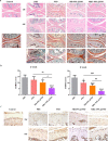
Histological analyses of different treatments for 2 or 6 weeks. a H&E (upper) and safranin-O/fast green staining (lower) of cartilage sections after the treatments with PBS, PSO, MR-PPL@PSO and MRC-PPL@PSO. Scale bar = 1 mm. b OARSI scores of articular cartilage after the treatments. Each data points represents mean ± S.D. (n = 5). *, # indicate p < 0.05; **, ## indicate p < 0.01; ***, ### indicate p < 0.001. c Immunohistochemical staining of MMP-13 on cartilage sections after the treatments. Scale bar = 300 μm

Similar articles

Cited by

References

    1. Dunlop DD, Manheim LM, Jing S, Chang RW. Arthritis prevalence and activity limitations in older adults. Arthritis Rheum. 2001;44:212–221. doi: 10.1002/1529-0131(200101)44:1<212::AID-ANR28>3.0.CO;2-Q. - DOI - PubMed
    1. Bender B, Perry M, Ramsey F, Boeselager G, Jackson-Thompson J. Prevalence and impact of chronic joint symptoms. J Am Med Assoc. 1998;279:1940–1941. doi: 10.1001/jama.279.24.1940. - DOI - PubMed
    1. Brown TJ, Laurent UB, Fraser: Turnover of hyaluronan in synovial joints: elimination of labelled hyaluronan from the knee joint of the rabbit. Experimental Physiology 1991, 76. - PubMed
    1. Albert C, Brocq O, Gerard D, Roux C, Euller-Ziegler L. Septic knee arthritis after intra-articular hyaluronate injection: two case reports. Joint Bone Spine. 2006;73:205–207. doi: 10.1016/j.jbspin.2005.03.005. - DOI - PubMed
    1. Bajpayee AG, Scheu M, Grodzinsky AJ, Porter RM. A rabbit model demonstrates the influence of cartilage thickness on intra-articular drug delivery and retention within cartilage. J Orthop Res. 2015;33:660–667. doi: 10.1002/jor.22841. - DOI - PubMed