MHC class II transactivator CIITA induces cell resistance to Ebola virus and SARS-like coronaviruses
- PMID: 32855215
- PMCID: PMC7665841
- DOI: 10.1126/science.abb3753
MHC class II transactivator CIITA induces cell resistance to Ebola virus and SARS-like coronaviruses
Abstract
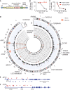
Recent outbreaks of Ebola virus (EBOV) and severe acute respiratory syndrome coronavirus 2 (SARS-CoV-2) have exposed our limited therapeutic options for such diseases and our poor understanding of the cellular mechanisms that block viral infections. Using a transposon-mediated gene-activation screen in human cells, we identify that the major histocompatibility complex (MHC) class II transactivator (CIITA) has antiviral activity against EBOV. CIITA induces resistance by activating expression of the p41 isoform of invariant chain CD74, which inhibits viral entry by blocking cathepsin-mediated processing of the Ebola glycoprotein. We further show that CD74 p41 can block the endosomal entry pathway of coronaviruses, including SARS-CoV-2. These data therefore implicate CIITA and CD74 in host defense against a range of viruses, and they identify an additional function of these proteins beyond their canonical roles in antigen presentation.
Copyright © 2020 The Authors, some rights reserved; exclusive licensee American Association for the Advancement of Science. No claim to original U.S. Government Works.
Figures


Comment in
-
Battle at the entrance gate: CIITA as a weapon to prevent the internalization of SARS-CoV-2 and Ebola viruses.Signal Transduct Target Ther. 2020 Nov 24;5(1):278. doi: 10.1038/s41392-020-00405-2. Signal Transduct Target Ther. 2020. PMID: 33235189 Free PMC article. No abstract available.
Similar articles
-
Newly Discovered Cellular Pathway Blocks Ebola, COVID-19 Viruses.JAMA. 2020 Nov 3;324(17):1712. doi: 10.1001/jama.2020.19649. JAMA. 2020. PMID: 33141192 No abstract available.
-
Ebola virus and severe acute respiratory syndrome coronavirus display late cell entry kinetics: evidence that transport to NPC1+ endolysosomes is a rate-defining step.J Virol. 2015 Mar;89(5):2931-43. doi: 10.1128/JVI.03398-14. Epub 2014 Dec 31. J Virol. 2015. PMID: 25552710 Free PMC article.
-
LY6E Restricts Entry of Human Coronaviruses, Including Currently Pandemic SARS-CoV-2.J Virol. 2020 Aug 31;94(18):e00562-20. doi: 10.1128/JVI.00562-20. Print 2020 Aug 31. J Virol. 2020. PMID: 32641482 Free PMC article.
-
The MHC Class II Transactivator CIITA: Not (Quite) the Odd-One-Out Anymore among NLR Proteins.Int J Mol Sci. 2021 Jan 22;22(3):1074. doi: 10.3390/ijms22031074. Int J Mol Sci. 2021. PMID: 33499042 Free PMC article. Review.
-
Remdesivir against COVID-19 and Other Viral Diseases.Clin Microbiol Rev. 2020 Oct 14;34(1):e00162-20. doi: 10.1128/CMR.00162-20. Print 2020 Dec 16. Clin Microbiol Rev. 2020. PMID: 33055231 Free PMC article. Review.
Cited by
-
CytoTalk: De novo construction of signal transduction networks using single-cell transcriptomic data.Sci Adv. 2021 Apr 14;7(16):eabf1356. doi: 10.1126/sciadv.abf1356. Print 2021 Apr. Sci Adv. 2021. PMID: 33853780 Free PMC article.
-
Integrating single-cell sequencing data with GWAS summary statistics reveals CD16+monocytes and memory CD8+T cells involved in severe COVID-19.Genome Med. 2022 Feb 17;14(1):16. doi: 10.1186/s13073-022-01021-1. Genome Med. 2022. PMID: 35172892 Free PMC article.
-
Structural aspects of the MHC expression control system.Biophys Chem. 2022 May;284:106781. doi: 10.1016/j.bpc.2022.106781. Epub 2022 Feb 15. Biophys Chem. 2022. PMID: 35228036 Free PMC article. Review.
-
Single-cell RNA-seq public data reveal the gene regulatory network landscape of respiratory epithelial and peripheral immune cells in COVID-19 patients.Front Immunol. 2023 Oct 23;14:1194614. doi: 10.3389/fimmu.2023.1194614. eCollection 2023. Front Immunol. 2023. PMID: 37936693 Free PMC article.
-
Mutants of human ACE2 differentially promote SARS-CoV and SARS-CoV-2 spike mediated infection.PLoS Pathog. 2021 Jul 16;17(7):e1009715. doi: 10.1371/journal.ppat.1009715. eCollection 2021 Jul. PLoS Pathog. 2021. PMID: 34270613 Free PMC article.
References
-
- Gire S. K., Goba A., Andersen K. G., Sealfon R. S. G., Park D. J., Kanneh L., Jalloh S., Momoh M., Fullah M., Dudas G., Wohl S., Moses L. M., Yozwiak N. L., Winnicki S., Matranga C. B., Malboeuf C. M., Qu J., Gladden A. D., Schaffner S. F., Yang X., Jiang P.-P., Nekoui M., Colubri A., Coomber M. R., Fonnie M., Moigboi A., Gbakie M., Kamara F. K., Tucker V., Konuwa E., Saffa S., Sellu J., Jalloh A. A., Kovoma A., Koninga J., Mustapha I., Kargbo K., Foday M., Yillah M., Kanneh F., Robert W., Massally J. L. B., Chapman S. B., Bochicchio J., Murphy C., Nusbaum C., Young S., Birren B. W., Grant D. S., Scheiffelin J. S., Lander E. S., Happi C., Gevao S. M., Gnirke A., Rambaut A., Garry R. F., Khan S. H., Sabeti P. C., Genomic surveillance elucidates Ebola virus origin and transmission during the 2014 outbreak. Science 345, 1369–1372 (2014). 10.1126/science.1259657 - DOI - PMC - PubMed
-
- Chen L., Stuart L., Ohsumi T. K., Burgess S., Varshney G. K., Dastur A., Borowsky M., Benes C., Lacy-Hulbert A., Schmidt E. V., Transposon activation mutagenesis as a screening tool for identifying resistance to cancer therapeutics. BMC Cancer 13, 93 (2013). 10.1186/1471-2407-13-93 - DOI - PMC - PubMed
-
- Bergemann T. L., Starr T. K., Yu H., Steinbach M., Erdmann J., Chen Y., Cormier R. T., Largaespada D. A., Silverstein K. A. T., New methods for finding common insertion sites and co-occurring common insertion sites in transposon- and virus-based genetic screens. Nucleic Acids Res. 40, 3822–3833 (2012). 10.1093/nar/gkr1295 - DOI - PMC - PubMed
-
- Carette J. E., Raaben M., Wong A. C., Herbert A. S., Obernosterer G., Mulherkar N., Kuehne A. I., Kranzusch P. J., Griffin A. M., Ruthel G., Dal Cin P., Dye J. M., Whelan S. P., Chandran K., Brummelkamp T. R., Ebola virus entry requires the cholesterol transporter Niemann-Pick C1. Nature 477, 340–343 (2011). 10.1038/nature10348 - DOI - PMC - PubMed
Publication types
MeSH terms
Substances
Grants and funding
LinkOut - more resources
Full Text Sources
Other Literature Sources
Medical
Molecular Biology Databases
Research Materials
Miscellaneous

