. 2020 Aug 27;11(1):4289.
doi: 10.1038/s41467-020-18039-x.

Senolytics prevent mt-DNA-induced inflammation and promote the survival of aged organs following transplantation

Affiliations

Senolytics prevent mt-DNA-induced inflammation and promote the survival of aged organs following transplantation

Jasper Iske et al. Nat Commun. .

Abstract

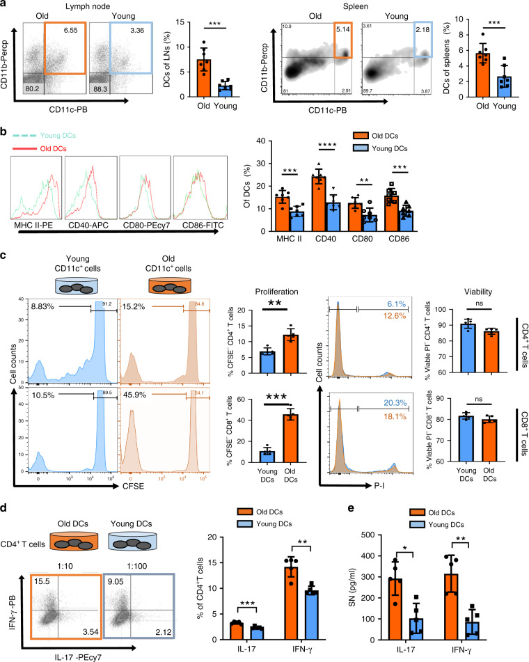
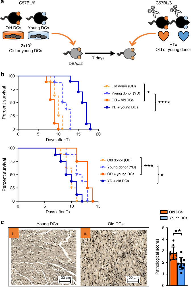
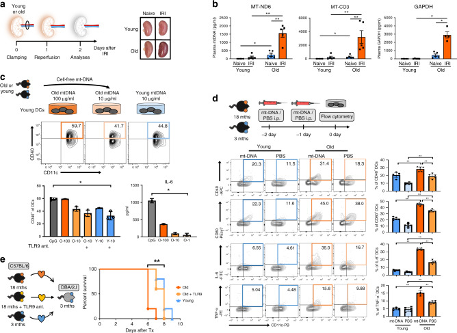
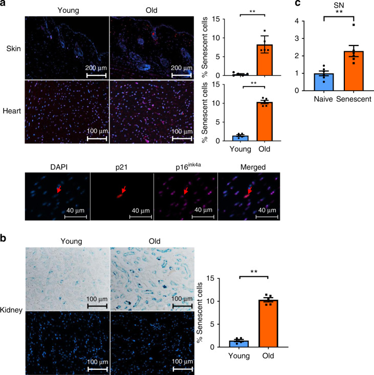
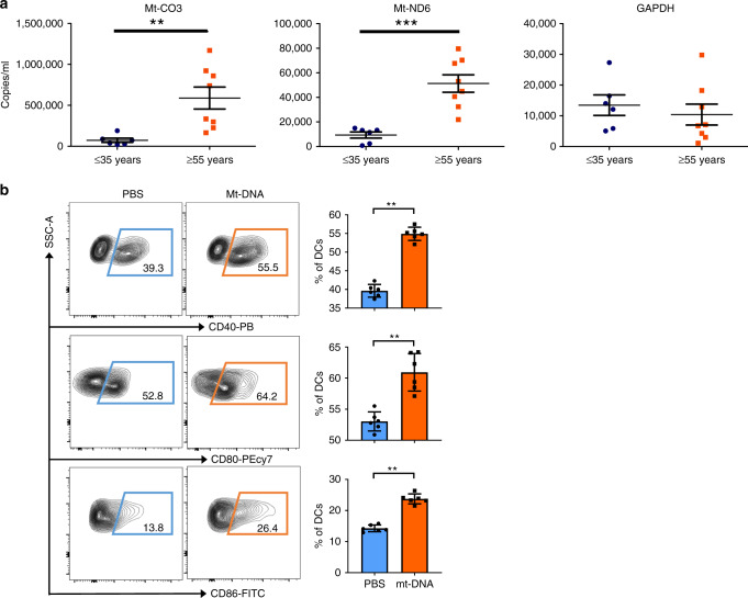
Older organs represent an untapped potential to close the gap between demand and supply in organ transplantation but are associated with age-specific responses to injury and increased immunogenicity, thereby aggravating transplant outcomes. Here we show that cell-free mitochondrial DNA (cf-mt-DNA) released by senescent cells accumulates with aging and augments immunogenicity. Ischemia reperfusion injury induces a systemic increase of cf-mt-DNA that promotes dendritic cell-mediated, age-specific inflammatory responses. Comparable events are observed clinically, with the levels of cf-mt-DNA elevated in older deceased organ donors, and with the isolated cf-mt-DNA capable of activating human dendritic cells. In experimental models, treatment of old donor animals with senolytics clear senescent cells and diminish cf-mt-DNA release, thereby dampening age-specific immune responses and prolonging the survival of old cardiac allografts comparable to young donor organs. Collectively, we identify accumulating cf-mt-DNA as a key factor in inflamm-aging and present senolytics as a potential approach to improve transplant outcomes and availability.


Conflict of interest statement

J.L.K. and T.T. have a financial interest related to this research. Patents on senolytic drugs are held by Mayo Clinic. This research has been reviewed by the Mayo Clinic Conflict of Interest Review Board and was conducted in compliance with Mayo Clinic Conflict of Interest policies. No conflicts of interest, financial or otherwise, are declared by the other authors.

Figures

Fig. 1
Fig. 1. Dendritic cells from old mice exhibit an activated phenotype and promote Th1 and Th17 T cell responses.
Single cell suspensions of lymph nodes and spleens from old and young C57BL/6 mice were labeled with anti-CD11c, anti-CD11b, anti-MHC class II, anti-CD40, anti-CD80, and anti-CD86. a The frequency of CD11b+CD11c+ DCs in lymph nodes (p = 0.0006) and spleens (p = 0.0023), and b the expression of costimulatory molecules (MHC-II: p = 0.0005/CD40: p < 0.0001/CD80: p = 0.0035/CD86: p = 0.0007) by splenic DCs were assessed by flow cytometry. c Proliferative capacities of CD4+ (p = 0.0079) and CD8+ T cells (p = 0.0079) co-cultured with DCs from old and young mice were determined using CFSE dilution and viability assessed using propidium iodide (d) CD4+ T cells from young mice were co-cultured with CD11b+CD11c+ DCs isolated from young and old mice and (e) pro-inflammatory cytokine expression was assessed by flow cytometry (IL-17: p = 0.0002/IFN-γ: p = 0.0013) and ELISA (Il-17: p = 0.004/IFN-γ: p = 0.0079); (n = 7 biologically independent animals), results are representative of at least three independent experiments. Column plots display mean with standard deviation. Statistical significance was determined by using two-tailed Mann–Whitney-test. Asterisks indicate p values *p ≤ 0.05, **p ≤ 0.01, and ***p ≤ 0.001, only significant values are shown. Source data are provided as a Source Data file.
Fig. 2
Fig. 2. Dendritic cells from old mice impair cardiac allograft survival in young DBA/2J mice.
a 2 × 106 CD11b+CD11c+ DCs were sorted from old and young C57BL/6 mice and administered i.v. into young DBA/2J mice 7 days prior to allogeneic cardiac transplants. b Kaplan–Meier analysis of cardiac allografts from old and young C57BL/6 mice transplanted into untreated DBA/2J recipients compared with those that had received adoptively transferred CD11b+CD11c+ DCs from old and young B6 mice (old donor vs. old donor + old DCs (p = 0.0216)/+ young DCs (p = 0.0005) and young donor vs. young donor + young DCs (p < 0.0001)/+ old DCs (0.0182); (n = 8 biologically independent animals). c By day 11 after transplantation, grafts were procured, perfused, and 5 mm sections were stained with H&E for pathological evaluation (p = 0.0011); n = 10/group, results are representative of at least three independent experiments. Column plots display mean with standard of the mean (SEM). Statistical significance for survival data was determined by log-rank Mantel–Cox test while pathological scores were compared by two-tailed Mann–Whitney-test. Asterisks indicate p values *p ≤ 0.05, **p ≤ 0.01, and ***p ≤ 0.001, only significant values are shown. Source data are provided as a Source Data file.
Fig. 3
Fig. 3. Systemic cf-mt-DNA increases upon IRI in old mice and promotes DC maturation through TLR9.
a Ischemia reperfusion injury was induced by clamping the renal pedicle of young and old C57Bl/6 (2 and 18 months) mice for 22 min, respectively. IRI and naive animals were euthanized after 48 h and kidneys were procured. The image shows the macroscopic appearance of kidneys directly after IRI. b Cell-free mitochondrial DNA (cf-mt-DNA) was quantified in plasma by real-time PCR according to standard curve results (young IRI vs. old IRI for mt-CO3: p = 0.0079); Column plots display Mean ± SEM; (n = 5 biologically independent animals). c Different concentrations of cf-mt-DNA isolated from young and old mice were added to DC cultured from young mice, costimulatory molecule expression was analyzed by flow cytometry with or in absence of a TLR9 antagonist (CpG vs. young + TLR9 ant: p = 0.0376), and Il-6 production was measured by ELISA (CpG vs. O1: p = 0.0134); statistical significance was determined using Kruskal–Wallis test with Dunn´s post hoc test; (n = 3 biologically independent samples). d Young or old C57BL/6 mice were injected i.v. with either 30 mg mt-DNA or PBS for 2 consecutive days. Subsequently, costimulatory molecule and cytokine expression of splenic DCs was analyzed by flow cytometry (young + mt-DNA vs. old + mt-DNA: (p = 0.0022); (n = 6 biologically independent animals). e Immediately after receiving a fully mismatched cardiac allograft from 18 months old C57Bl/6 donor mouse, 3 months old DBA2/J recipient mice were treated every 24 h with an i.p. TLR9 antagonist for the follow-up period (p = 0.0009), (n = 5 biologically independent animals); results are representative of at least three independent experiments. Column plots display mean with standard deviation. Statistical significance was determined using two-tailed Mann–Whitney test. Survival was compared by log-rank Mantel–Cox test. Asterisks indicate p values *p ≤ 0.05, **p ≤ 0.01, and ***p ≤ 0.001, only significant values are shown. Source data are provided as a Source Data file.
Fig. 4
Fig. 4. Senescent cells accumulate with aging and are a source of cf-mt-DNA with aging.
Skin, hearts, and kidneys were procured from old and young C57BL/6 mice and embedded in paraffin. a Skin and hearts were cut into slides and co-stained for p16Ink4a, p21Cip1, and DAPI; b frozen slides of kidneys were made and subsequently stained for sa-β-gal. The percentage of senescent cells was defined as the number of (a) p16/p21 double-positive cells or (b) sa-β-gal-positive cells of DAPI-stained cells using a confocal microscope (p = 0.0079), (n = 5 biologically independent animals). c Mouse adipocytes were isolated from C57BL/6 mice and senescence induced using 30 serial passages or 10 Gy irradiation. Cf-mt-DNA levels were measured in supernatants by real-time PCR comparing senescent and naive cell cultures (p = 0.0022); (n = 6 biologcially independent samples); column plots display mean ± SD, results are representative of at least three independent experiments. Statistical significance was determined by using two-tailed Mann–Whitney test. Asterisks indicate p values *p ≤ 0.05, **p ≤ 0.01, and ***p ≤ 0.001, ****p ≤ 0.0001, only significant values are shown. Source data are provided as a Source Data file.
Fig. 5
Fig. 5. Old human organ donors had increased systemic levels of cf-mt-DNA activating DCs.
a Cf-mt-DNA was quantified in the plasma of young (<35 years) and old (>55 years) organ donors by RT-PCR using TAQMAN primers for mt-Co3 (p = 0.0013) and mt-nd6 (p = 0.0007), Scatter plots display mean ± SEM. b Dendritic cells were differentiated from isolated PBMC, stimulated with human mt-DNA (10 μg/ml), and costimulatory molecule expression was analyzed using flow cytometry (p = 0.0022). Column plots display mean ± SD (n = 6 biologically independent samples); results are representative of at least three independent experiments. Statistical significance was determined by using two-tailed Mann–Whitney-test. Asterisks indicate p values *p ≤ 0.05, **p ≤ 0.01, and ***p ≤ 0.001, only significant values are shown. Source data are provided as a Source Data file.
Fig. 6
Fig. 6. Senolytics decrease the number of senescent cells, reduce cf-mt-DNA levels, alleviate systemic inflammatory immune response after IRI, and prolong cardiac allograft survival.
a Old C57/B6 mice were treated with senolytics (D and Q, or either D or Q in b) on 3 successive days/week. After 1 month, kidney, heart, and skin were procured, stained, and the percentage of senescent cells assessed as described in Fig. 4, (p = 0.0079); Column plots display mean ± SD, (n = 5 biologically independent animals). b Systemic levels of p16Ink4a and cf-mt-DNA were measured by real-time PCR and calculated relative to GAPDH expression (old vs. old + D and Q: p = 0.0317); Column plots display mean ± SEM, (n = 6 biologically independent animals). c Young and old C57BL/6 mice were treated with senolytics for 3 successive days/week for 1 month. Subsequently, IRI was induced in young and old animals; IL-17 and IFN-γ expression of CD4+ and CD8+ T cells were assessed by flow cytometry (old control vs. old treated: CD4+IL-17+ (p = 0.0087)/CD4+IFN-γ+ (p = 0.0022)/CD8+IFN-γ+ (p = 0.0022); column plots display mean ± SD, (n = 6 biologically independent animals). d Old and young donor C57BL/6 mice were treated with D and Q or PBS prior to fully mismatched cardiac transplantation and allograft survival was monitored by daily palpation (p = 0.0012); (n = 8 biologically independent animals). e Old and young donor C57BL/6 mice were treated with D and Q or PBS prior to fully mismatched cardiac transplantation while recipient animals were treated with weekly injections of CTLA4-IG following transplantation and allograft survival was monitored by daily palpation (p = 0.0064); (n = 5 biologically independent animals). Results are representative of at least three independent experiments. Statistical significance was determined by two-tailed Mann–Whitney test. Survival was compared by log-rank Mantel–Cox test. Asterisks indicate p values *p ≤ 0.05, **p ≤ 0.01, and ***p ≤ 0.001, ****p ≤ 0.0001, only significant values are shown. Source data are provided as a Source Data file.

Comment in

References

    1. Korolchuk VI, Miwa S, Carroll B, von Zglinicki T. Mitochondria in cell senescence: is mitophagy the weakest link? EBioMedicine. 2017;21:7–13. - PMC - PubMed
    1. Tullius SG, Rabb H. Improving the supply and quality of deceased-donor organs for transplantation. N. Engl. J. Med. 2018;378:1920–1929. - PubMed
    1. Klassen DK, et al. The OPTN deceased donor potential study: implications for policy and practice. Am. J. Transpl. 2016;16:1707–1714. - PubMed
    1. Tullius SG, Milford E. Kidney allocation and the aging immune response. N. Engl. J. Med. 2011;364:1369–1370. - PubMed
    1. Oberhuber R, et al. CD11c+ dendritic cells accelerate the rejection of older cardiac transplants via interleukin-17A. Circulation. 2015;132:122–131. - PMC - PubMed

Publication types

MeSH terms