

Comparison of physical examination, ultrasound techniques and magnetic resonance imaging for the diagnosis of deep infiltrating endometriosis: A systematic review and meta-analysis of diagnostic accuracy studies
- PMID: 32855690
- PMCID: PMC7444323
- DOI: 10.3892/etm.2020.9043
Comparison of physical examination, ultrasound techniques and magnetic resonance imaging for the diagnosis of deep infiltrating endometriosis: A systematic review and meta-analysis of diagnostic accuracy studies
Abstract
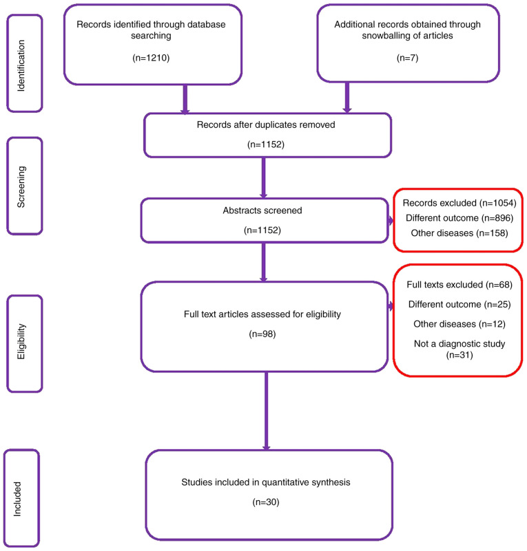
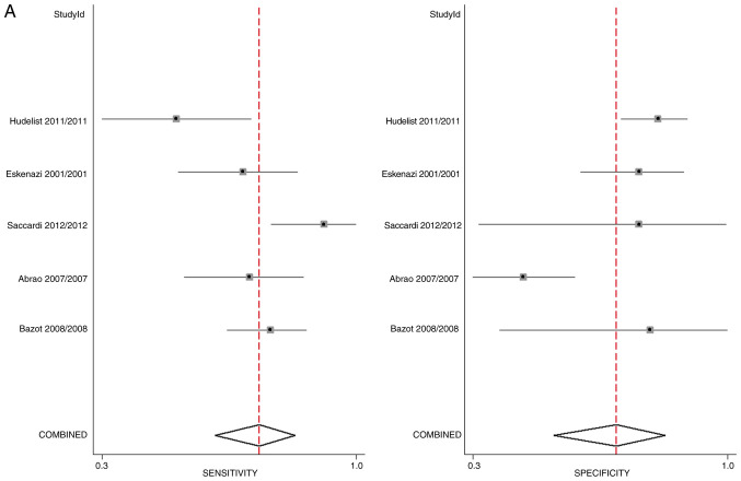
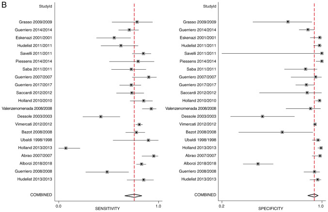
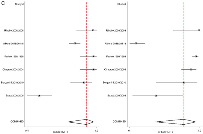
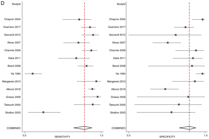
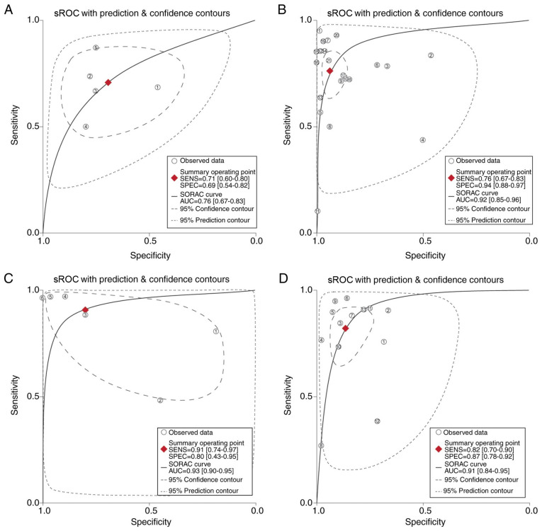
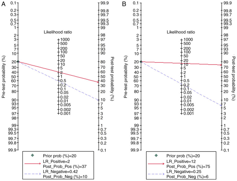
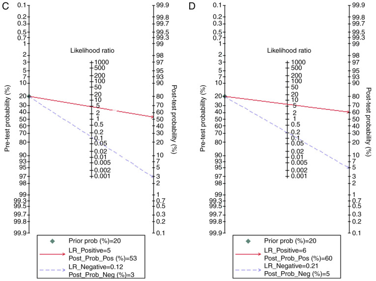
Visual inspection via laparoscopy is considered the gold standard for the diagnosis of deep infiltrating endometriosis. Laparoscopy is an invasive procedure; therefore, it would be beneficial to patients if accurate non-invasive modalities were available for the diagnosis of deep infiltrating endometriosis. The purpose of the current review and meta-analysis was to assess the diagnostic accuracy of clinical examination, transvaginal ultrasound (TVUS), transrectal ultrasound (TRUS) and MRI as alternative methods for diagnosis of deep infiltrating endometriosis. A systematic search of the Medline, Scopus, Embase and Cochrane library databases, between their inception and September 2019, was performed. The quality of trials was assessed using the quality assessment of diagnostic accuracy studies-2 tool. Meta-analyses were conducted to obtain the pooled sensitivity, specificity, positive and negative likelihood ratios and diagnostic odds ratio for each of the three imaging modalities and clinical examination. A total of 30 studies with 4,565 participants were included in the review. Physical examination had a pooled sensitivity of 71% and a specificity of 69%, with an average diagnostic accuracy [area under the curve (AUC) =0.76]. TVUS had a pooled sensitivity of 76% and a specificity of 94%, with higher diagnostic accuracy than physical examination (AUC =0.92). TRUS had a pooled sensitivity of 91% and a specificity of 80% with an AUC of 0.93. MRI had a pooled sensitivity of 82% and a specificity of 87% with higher diagnostic accuracy than physical examination (AUC =0.91). All the imaging modalities had good clinical utility, as indicated by the Fagan plot. The present analysis demonstrates that the imaging modalities TVUS, TRUS and MRI may be highly useful alternatives to laparoscopy for diagnosis of deep infiltrating endometriosis and that these techniques have a high sensitivity and specificity.
Keywords: MRI; deep infiltrating endometriosis; ultrasonography; validation.
Copyright: © Zhang et al.
Figures
Similar articles
-
Systematic evaluation of endometriosis by transvaginal ultrasound can accurately replace diagnostic laparoscopy, mainly for deep and ovarian endometriosis.Hum Reprod. 2021 May 17;36(6):1492-1500. doi: 10.1093/humrep/deab085. Hum Reprod. 2021. PMID: 33864088
-
Correlation between ultrasound findings and laparoscopy in prediction of deep infiltrating endometriosis (DIE).Aust N Z J Obstet Gynaecol. 2020 Dec;60(6):946-951. doi: 10.1111/ajo.13242. Epub 2020 Sep 7. Aust N Z J Obstet Gynaecol. 2020. PMID: 32895927
-
Noninvasive Diagnosis of Adenomyosis: A Structured Review and Meta-analysis of Diagnostic Accuracy in Imaging.J Minim Invasive Gynecol. 2020 Feb;27(2):408-418.e3. doi: 10.1016/j.jmig.2019.11.001. Epub 2019 Nov 8. J Minim Invasive Gynecol. 2020. PMID: 31712162
-
[Meta-analysis of ultrasonography in diagnosis of deeply infiltrating endometriosis].Zhonghua Fu Chan Ke Za Zhi. 2010 Apr;45(4):269-72. Zhonghua Fu Chan Ke Za Zhi. 2010. PMID: 20646538 Chinese.
-
Can Enhanced Techniques Improve the Diagnostic Accuracy of Transvaginal Sonography and Magnetic Resonance Imaging for Rectosigmoid Endometriosis? A Systematic Review and Meta-analysis.J Obstet Gynaecol Can. 2020 Apr;42(4):488-499.e4. doi: 10.1016/j.jogc.2019.07.016. Epub 2019 Nov 23. J Obstet Gynaecol Can. 2020. PMID: 31767378
Cited by
-
The experiences of endometriosis patients with diagnosis and treatment in New Zealand.Front Glob Womens Health. 2022 Aug 31;3:991045. doi: 10.3389/fgwh.2022.991045. eCollection 2022. Front Glob Womens Health. 2022. PMID: 36118149 Free PMC article.
-
MRI of endometriosis in correlation with the #Enzian classification: applicability and structured report.Insights Imaging. 2023 Jul 5;14(1):120. doi: 10.1186/s13244-023-01466-x. Insights Imaging. 2023. PMID: 37405519 Free PMC article. Review.
-
Vault Endometriosis: Detailed Step-by-Step Laparoscopic Surgical Management Technique.JSLS. 2021 Oct-Dec;25(4):e2021.00057. doi: 10.4293/JSLS.2021.00057. JSLS. 2021. PMID: 34803369 Free PMC article.
-
Society of Radiologists in Ultrasound Consensus on Routine Pelvic US for Endometriosis.Radiology. 2024 Apr;311(1):e232191. doi: 10.1148/radiol.232191. Radiology. 2024. PMID: 38591980 Free PMC article. Review.
-
Agreement between magnetic resonance imaging and ultrasonography in deep pelvic endometriosis.Rev Assoc Med Bras (1992). 2025 Mar 31;71(2):e20241235. doi: 10.1590/1806-9282.20241235. eCollection 2025. Rev Assoc Med Bras (1992). 2025. PMID: 40172392 Free PMC article.
References
-
- Irving JA, Clement PB. Diseases of the Peritoneum. In: Blaustein's Pathology of the Female Genital Tract. Kurman RJ, Ellenson LH and Ronnett BM (eds) Springer US, Boston, MA, pp625-678, 2011.
-
- Wheeler JM. Epidemiology of endometriosis-associated infertility. J Reprod Med. 1989;34:41–46. - PubMed
LinkOut - more resources
Full Text Sources