. 2020 Mar 5:95:381-414.
doi: 10.1016/j.simyco.2020.02.004. eCollection 2020 Mar.

Evolution of lifestyles in Capnodiales

Affiliations

Evolution of lifestyles in Capnodiales

J Abdollahzadeh et al. Stud Mycol. .

Abstract

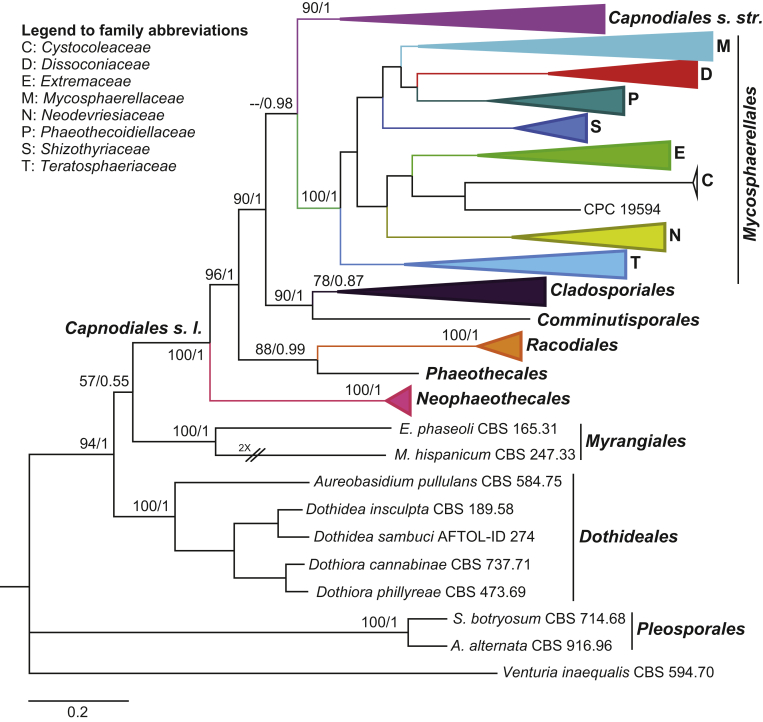
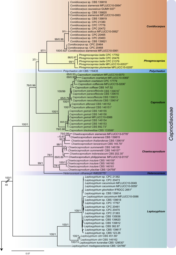
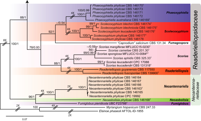
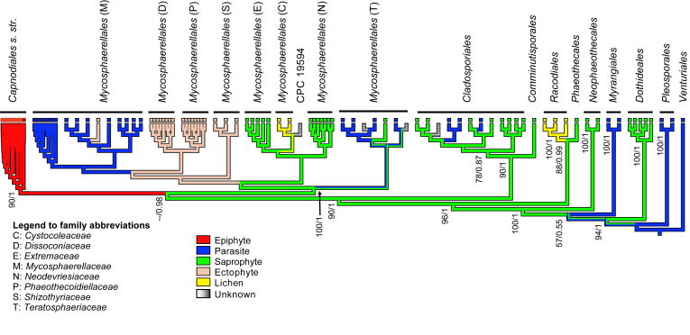
The Capnodiales, which includes fungi known as the sooty moulds, represents the second largest order in Dothideomycetes, encompassing morphologically and ecologically diverse fungi with different lifestyles and modes of nutrition. They include saprobes, plant and human pathogens, mycoparasites, rock-inhabiting fungi (RIF), lichenised, epi-, ecto- and endophytes. The aim of this study was to elucidate the lifestyles and evolutionary patterns of the Capnodiales as well as to reconsider their phylogeny by including numerous new collections of sooty moulds, and using four nuclear loci, LSU, ITS, TEF-1α and RPB2. Based on the phylogenetic results, combined with morphology and ecology, Capnodiales s. lat. is shown to be polyphyletic, representing seven different orders. The sooty moulds are restricted to Capnodiales s. str., while Mycosphaerellales is resurrected, and five new orders including Cladosporiales, Comminutisporales, Neophaeothecales, Phaeothecales and Racodiales are introduced. Four families, three genera, 21 species and five combinations are introduced as new. Furthermore, ancestral reconstruction analysis revealed that the saprobic lifestyle is a primitive state in Capnodiales s. lat., and that several transitions have occurred to evolve lichenised, plant and human parasitic, ectophytic (sooty blotch and flyspeck) and more recently epiphytic (sooty mould) lifestyles.

Keywords: Capnodiales; Capnodium alfenasii Abdollahz. & Crous; Capnodium blackwelliae Abdollahz. & Crous; Capnodium gamsii Abdollahz. & Crous; Capnodium neocoffeicola Abdollahz. & Crous; Capnodium paracoffeicola Abdollahz. & Crous; Chaetocapnodium indonesiacum Abdollahz. & Crous; Chaetocapnodium insulare Abdollahz. & Crous; Chaetocapnodium philippinense (Hongsanan & K.D. Hyde) Abdollahz. & Crous; Chaetocapnodium placitae (Cheewangkoon & Crous) Abdollahz. & Crous; Chaetocapnodium summerellii Abdollahz. & Crous; Chaetocapnodium tanzanicum Abdollahz. & Crous; Chaetocapnodium thailandense Abdollahz. & Crous; Cladosporiales Abdollahz. & Crous; Cladosporium; Comminutisporaceae Abdollahz. & Crous; Comminutisporales Abdollahz. & Crous; Leptoxyphium citri Abdollahz. & Crous; Multigene phylogeny; Mycosphaerella; Neoantennariella Abdollahz. & Crous; Neoantennariella phylicae Abdollahz. & Crous; Neoantennariellaceae Abdollahz. & Crous; Neoasbolisia Abdollahz. & Crous; Neoasbolisia phylicae Abdollahz. & Crous; Neophaeotheca Abdollahz. & Crous; Neophaeotheca salicorniae (Crous & Roets) Abdollahz. & Crous; Neophaeotheca triangularis (de Hoog & Beguin) Abdollahz. & Crous; Neophaeothecaceae Abdollahz. & Crous; Neophaeothecales Abdollahz. & Crous; Phaeothecales Abdollahz. & Crous; Phaeoxyphiella australiana Abdollahz. & Crous; Phaeoxyphiella phylicae Abdollahz. & Crous; Phragmocapnias plumeriae (Hongsanan & K.D. Hyde) Abdollahz. & Crous; Racodiales Abdollahz. & Crous; Readerielliopsidaceae Abdollahz. & Crous; Scolecoxyphium blechni Abdollahz. & Crous; Scolecoxyphium blechnicola Abdollahz. & Crous; Scolecoxyphium leucadendri Abdollahz. & Crous; Scolecoxyphium phylicae Abdollahz. & Crous; Scorias aphidis Abdollahz. & Crous; Scorias camelliae Abdollahz. & Crous; Sooty moulds.


Figures

Fig. 1
Fig. 1
Reduced phylogenetic tree inferred from a Bayesian analysis based on a concatenated alignment of LSU, TEF-1α and RPB2. Bayesian posterior probabilities (PP) and maximum likelihood bootstrap support values (ML-BS) are indicated at the nodes (PP/ML-BS). The scale bar represents the expected number of changes per site. The lineages in Capnodiales s. l. are indicated in different colours. The tree was rooted with Venturia inaequalis (CBS 594.70).
Fig. 2
Fig. 2
Phylogenetic tree inferred from a RAxML search of a concatenated alignment of LSU, ITS, TEF-1α and RPB2. Maximum likelihood bootstrap support values (ML-BS) and Bayesian posterior probabilities (PP) are indicated at the nodes (ML-BS/PP). The scale bar represents the expected number of changes per site. Families and orders are highlighted in blocks of different colour and indicated to the right of the tree. The tree was rooted with Myriangium hispanicum (CBS 247.33) and Elsinoe phaseoli (AFTOL-ID-1855). T Ex-type, ET Ex-epitype.
Fig. 2
Fig. 2
Phylogenetic tree inferred from a RAxML search of a concatenated alignment of LSU, ITS, TEF-1α and RPB2. Maximum likelihood bootstrap support values (ML-BS) and Bayesian posterior probabilities (PP) are indicated at the nodes (ML-BS/PP). The scale bar represents the expected number of changes per site. Families and orders are highlighted in blocks of different colour and indicated to the right of the tree. The tree was rooted with Myriangium hispanicum (CBS 247.33) and Elsinoe phaseoli (AFTOL-ID-1855). T Ex-type, ET Ex-epitype.
Fig. 3
Fig. 3
Cladogram showing the ancestral state reconstruction and evolution of lifestyles over the tree. Maximum likelihood bootstrap support values (ML-BS) and Bayesian posterior probabilities (PP) are indicated at the nodes (ML-BS/PP).
Fig. 4
Fig. 4
Capnodium alfenasii. A, B. Colony (2-wk-old) on MEA. C. Conidiomata arising from mycelia or immature conidiomata on SNA. D. Conidioma on SNA. E. Ostiole surround by hyaline hyphae. F. Conidia produced in ellipsoidal central part of conidioma. G. Conidia. Scale bars: C = 25 μm; D, E = 20 μm; F, G = 10 μm.
Fig. 5
Fig. 5
Capnodium blackwelliae. A, B. Colony (2-wk-old) on MEA. C–G. Conidiomata on SNA. H. Conidia. Scale bars: C–G = 25 μm; H = 10 μm.
Fig. 6
Fig. 6
Capnodium gamsii. A, B. Colony (2-wk-old) on MEA. C, D. Conidiomata arising from mycelia or immature conidiomata on SNA. E–H. Conidiomata on SNA. I. Conidia. Scale bars: C–F = 25 μm; G, H = 50 μm; I = 10 μm.
Fig. 7
Fig. 7
Capnodium neocoffeicola. A, B. Colony (2-wk-old) on MEA. C. Conidiomata arising from mycelia or immature conidiomata on SNA. D. Conidiomata on SNA. E. Conidia produced in ellipsoidal central part of conidioma. F. Conidia. Scale bars: C, E = 20 μm; D = 25 μm; F = 10 μm.
Fig. 8
Fig. 8
Capnodium paracoffeicola. A, B. Colony (2-wk-old) on MEA. C. Conidiomata arising from mycelia or immature conidiomata on SNA. D. Conidiomata on SNA. E. Ostiole surround by hyaline hyphae. F. Conidia. Scale bars: C–E = 25 μm; F = 10 μm.
Fig. 9
Fig. 9
Chaetocapnodium indonesiacum. A, B. Colony (2-wk-old) on MEA. C. Conidiomata on SNA. D. Conidia. Scale bars: C = 25 μm; D = 10 μm.
Fig. 10
Fig. 10
Chaetocapnodium insulare. A, B. Colony (2-wk-old) on MEA. C, D. Septate hyphae with mucilaginous outer wall layer and immature conidiomata on SNA. E. Conidiomata with setae. F. Conidia inside conidioma. G. Conidia. H. Ascoma with 3-septate brown ascospores. Scale bars: C–F, H = 20 μm; G = 10 μm.
Fig. 11
Fig. 11
Chaetocapnodium summerellii. A, B. Colony (2-wk-old) on MEA. C. Conidiomata on SNA. D. Conidioma with setae. E. Conidia inside conidiomata. F. Conidia. Scale bars: C, D = 25 μm; E = 20 μm; F = 10 μm.
Fig. 12
Fig. 12
Chaetocapnodium tanzanicum. A, B. Colony (2-wk-old) on MEA. C, D. Conidia inside conidiomata. E, F. Conidiomata with setae on SNA. G. Conidia. Scale bars: C, D, F = 25 μm; E = 50 μm; G = 10 μm.
Fig. 13
Fig. 13
Chaetocapnodium thailandense. A, B. Colony (2-wk-old) on MEA. C, D. Conidiomata with setae on SNA. E. Conidia inside conidioma. F. Conidia. Scale bars: C = 25 μm; D, E = 20 μm; F = 10 μm.
Fig. 14
Fig. 14
Conidiocarpus, Phragmocapnias and Polychaeton conidiomata. A, B.Conidiocarpus conidiomata on SNA. C–E.Conidiocarpus conidiomata on OA. F.Polychaeton conidioma on SNA. G, H.Polychaeton conidiomata on OA. I, J.Phragmocapnias conidiomata on SNA. Scale bars: A, C–E, G, H = 50 μm; F, J = 25 μm; I = 20 μm.
Fig. 15
Fig. 15
Leptoxyphium citri. A, B. Colony (2-wk-old) on MEA. C, D. Conidiomata arising from mycelia on SNA. E. Mature funnel-shaped conidioma at apex with hyaline hyphae surrounding the ostiole. F. Proliferation through the fertile head of conidioma. G. Conidia. H–J. Synasexual morph 2-celled conidia. Scale bars: C = 40 μm; D = 20 μm; E = 25 μm; F = 50 μm; G–J = 10 μm.
Fig. 16
Fig. 16
Phragmocapnias betle. A. Ascoma with setae on SNA. B. Asci and ascospores. C. Conidia. D. Conidioma arising from mycelium on SNA. E. Conidioma on SNA. Scale bars: A, D = 25 μm; E = 20 μm; B, C = 10 μm.
Fig. 17
Fig. 17
Neoantennariella phylicae. A, B. Colony (2-wk-old) on MEA. C. Septate hyphae and immature conidioma on SNA. D–F. Intercalary, lateral and terminal conidiomata. G. Conidia. Scale bars: C–D = 20 μm; E–F = 25 μm; G = 10 μm.
Fig. 18
Fig. 18
Neoasbolisia phylicae. A, B. Colony (2-wk-old) on MEA. C–E. Conidiomata on SNA. F. Conidia. Scale bars: C = 20 μm; D–E = 25 μm; F = 10 μm.
Fig. 19
Fig. 19
Phaeoxyphiella phylicae. A, B. Colony (2-wk-old) on MEA. C, D. Conidiomata with conidia on SNA. E. Transversely euseptate brown conidia. F, G. Spermogonia. H. Microconidia. Scale bars: C, E, F = 20 μm; D = 50 μm; G = 25 μm; H = 10 μm.
Fig. 20
Fig. 20
Scolecoxyphium blechni. A, B. Colony (2-wk-old) on MEA. C–H. Irregularly cylindrical-oblong, straight or flexuous, simple or branched conidiomata on SNA. I. Conidia. Scale bars: C = 50 μm; D, F–H = 20 μm; E = 25 μm; I = 10 μm.
Fig. 21
Fig. 21
Scolecoxyphium leucadendri. A, B. Colony (2-wk-old) on MEA. C. Immature conidiomata. D–F. Irregularly cylindrical-oblong, straight or flexuous, simple or branched conidiomata on SNA. G. Conidia inside conidiomata. H. Conidia. Scale bars: C, E = 20 μm; D, F = 25 μm; G, H = 10 μm.
Fig. 22
Fig. 22
Scolecoxyphium phylicae. A, B. Colony (2-wk-old) on MEA. C–F. Cylindrical-oblong, straight, simple conidiomata on SNA. G. Conidia. Scale bars: C = 25 μm; D–F = 20 μm; G = 10 μm.
Fig. 23
Fig. 23
Scorias aphidis. A, B. Colony (2-wk-old) on MEA. C, D. Flask-shape conidiomata on SNA. E. Ostioles surround by hyaline hyphae. F, G. Conidia produced in ellipsoidal part of conidiomata. H. Conidia. Scale bars: D = 50 μm; E–F = 25 μm; G–H = 10 μm.
Fig. 24
Fig. 24
Scorias camelliae. A, B. Colony (2-wk-old) on MEA. C. Flask-shape conidiomata on SNA. D. Ostiole surround by hyaline hyphae. E. Conidia. Scale bars: C–D = 20 μm; E = 10 μm.

References

    1. Ametrano C.G., Grewe F., Crous P.W., et al. Genome-scale data resolve ancestral rock-inhabiting lifestyle in Dothideomycetes (Ascomycota) IMA Fungus. 2019;10:19. - PMC - PubMed
    1. Aptroot A. CBS-KNAW Fungal Biodiversity Centre; Utrecht, The Netherlands: 2006. Mycosphaerella and its anamorphs 2. Conspectus of mycosphaerella; pp. 1–231. CBS Biodiversity Series 5.
    1. Batista A.C., Ciferri R. Capnodiales. Saccardoa. 1963;2:1–296.
    1. Batista A.C., Ciferri R. The sooty-molds of the family Asbolisiaceae. Quaderno del Laboratorio Crittogamico del Istituto Botanico dell'Università di Pavia. 1963;31:1–229.
    1. Bensch K., Braun U., Groenewald J.Z., et al. The genus Cladosporium. Studies in Mycology. 2012;72:1–401. - PMC - PubMed