. 2020 Jun 29:9:656.
doi: 10.12688/f1000research.24680.1. eCollection 2020.

Isolation and characterization of a novel bacterial strain from a Tris-Acetate-Phosphate agar medium plate of the green micro-alga Chlamydomonas reinhardtii that can utilize common environmental pollutants as a carbon source

Affiliations

Isolation and characterization of a novel bacterial strain from a Tris-Acetate-Phosphate agar medium plate of the green micro-alga Chlamydomonas reinhardtii that can utilize common environmental pollutants as a carbon source

Mautusi Mitra et al. F1000Res. .

Abstract

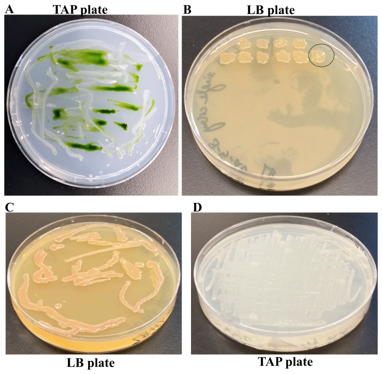
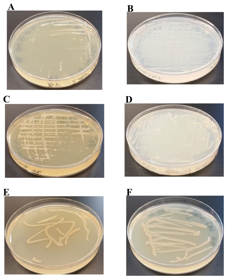
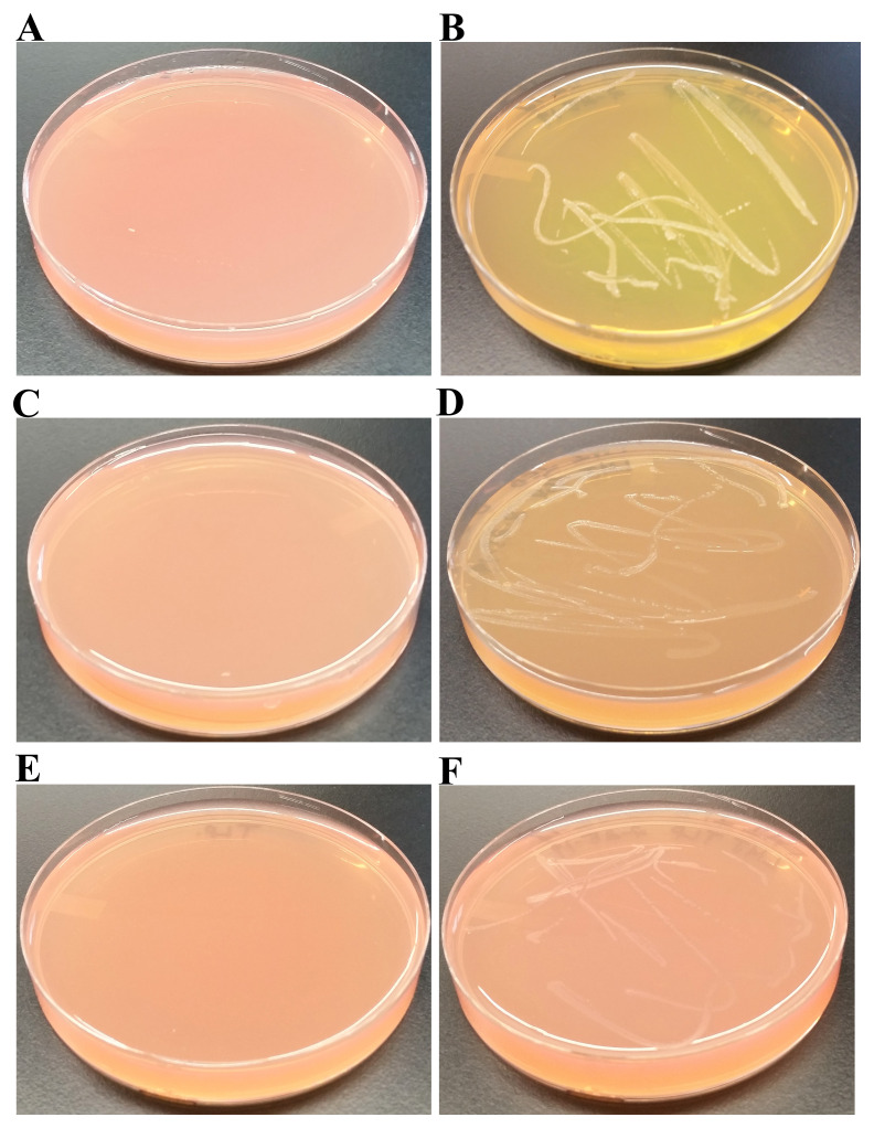
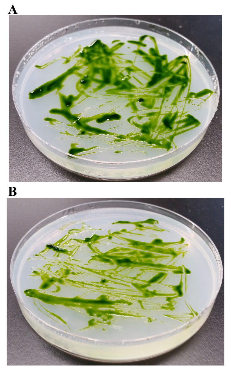
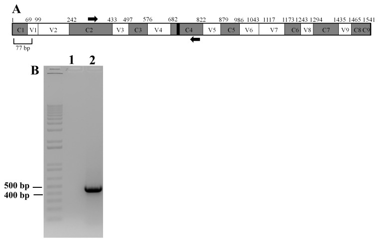
Background:Chlamydomonas reinhardtii, a green micro-alga can be grown at the lab heterotrophically or photo-heterotrophically in Tris-Phosphate-Acetate (TAP) medium which contains acetate as the sole carbon source. When grown in TAP medium, Chlamydomonas can utilize the exogenous acetate in the medium for gluconeogenesis using the glyoxylate cycle, which is also present in many bacteria and higher plants. A novel bacterial strain, LMJ, was isolated from a contaminated TAP medium plate of Chlamydomonas. We present our work on the isolation and physiological and biochemical characterizations of LMJ. Methods: Several microbiological tests were conducted to characterize LMJ, including its sensitivity to four antibiotics. We amplified and sequenced partially the 16S rRNA gene of LMJ. We tested if LMJ can utilize cyclic alkanes, aromatic hydrocarbons, poly-hydroxyalkanoates, and fresh and combusted car motor oil as the sole carbon source on Tris-Phosphate (TP) agar medium plates for growth. Results: LMJ is a gram-negative rod, oxidase-positive, mesophilic, non-enteric, pigmented, salt-sensitive bacterium. LMJ can ferment glucose, is starch hydrolysis-negative, and is very sensitive to penicillin and chloramphenicol. Preliminary spectrophotometric analyses indicate LMJ produces pyomelanin. NCBI-BLAST analyses of the partial 16S rRNA gene sequence of LMJ showed that it matched to that of an uncultured bacterium clone LIB091_C05_1243. The nearest genus relative of LMJ is an Acidovorax sp. strain. LMJ was able to use alkane hydrocarbons, fresh and combusted car motor oil, poly-hydroxybutyrate, phenanthrene, naphthalene, benzoic acid and phenyl acetate as the sole carbon source for growth on TP-agar medium plates. Conclusions: LMJ has 99.14% sequence identity with the Acidovorax sp. strain A16OP12 whose genome has not been sequenced yet. LMJ's ability to use chemicals that are common environmental pollutants makes it a promising candidate for further investigation for its use in bioremediation and, provides us with an incentive to sequence its genome.

Keywords: 16S rRNA gene; Acidovorax sp.; Chlamydomonas; LMJ; TAP medium; bioremediation; penicillin-sensitivity; pyomelanin.


Conflict of interest statement

No competing interests were disclosed.

Figures

Figure 1.
Figure 1.. Isolation of a novel bacterial strain from a contaminated Chlamydomonas Tris-Acetate-Phosphate (TAP)-agar medium plate.
( A) TAP-agar medium plate showing bacterial contamination of a Chlamydomonas strain, LMJ.SG0182 at room temperature (22ºC). The bacterium is named LMJ, after the abbreviated Chlamydomonas LMJ.SG0182 strain. ( B) Ten single colonies of LMJ on a lysogeny broth (LB)-agar medium plate. Colony # 10 outlined by the black circle was picked for culture stock maintenance and further analyses. ( C) LB-agar medium plate of purified LMJ strain ( D) TAP-agar medium plate of purified LMJ strain. Culture plates shown in ( C) and ( D) were imaged after 5 days of growth at 37ºC.
Figure 2.
Figure 2.. Gram-stained LMJ imaged under 100X magnification.
LMJ cells from a LB-agar medium plate was used for gram-staining. Gram-stained cells were visualized and imaged under an oil immersion lens of a bright-field microscope.
Figure 3.
Figure 3.. LMJ growth on TAP- and LB-agar medium plates at different temperatures.
( A) Growth on LB-agar medium plate at room temperature (22ºC). ( B) Growth on TAP-agar medium plate at room temperature. ( C) Growth on LB-agar medium plate at 37ºC. ( D) Growth on TAP-agar medium plate at 37ºC. ( E) LMJ growth on Mueller-Hinton agar medium plate at 22ºC. ( F) LMJ growth on Mueller-Hinton agar medium plate at 37ºC. Culture plates were imaged after 5 days of growth.
Figure 4.
Figure 4.. Effect of 1% NaCl on LMJ growth at different temperatures.
( A) LMJ growth on LB (contains 1% NaCl)-agar medium plate at 22ºC. ( B) LMJ growth on LB (minus 1% NaCl)-agar medium plate at 22ºC. ( C) LMJ growth on LB (contains 1% NaCl)-agar medium plate at 37ºC. ( D) LMJ growth on LB (minus 1% NaCl)-agar medium plate at 37ºC. ( E) LMJ growth on TAP+1% NaCl-agar medium plate at 22ºC. ( F) LMJ growth on TAP-agar medium plate at 22ºC. ( G) LMJ growth on TAP+1% NaCl-agar medium plate at 37ºC. ( H) LMJ growth on TAP-agar medium plate at 37ºC. Culture plates were imaged after 3 days of growth.
Figure 5.
Figure 5.. Liquid medium grown-LMJ cells are prone to cell lysis during Gram staining.
( A) A culture of LMJ grown for 24 h in liquid TAP and liquid TAP medium (control). ( B) Gram-stained LMJ cells from the 24 hours-TAP liquid culture. ( C) A 24 hours-grown culture of LMJ in liquid LB and liquid LB medium (control). ( D) Gram-stained LMJ cells from the 24 hours-grown LMJ LB liquid culture. Mostly membrane debris, few rods and round shaped cells can be seen. Original gram-stained files can be found at 10.6084/m9.figshare.12420893.
Figure 6.
Figure 6.. Pigment production in LMJ in dark and in light on TAP + 1% Tryptone-agar.
( A) LMJ on TAP +1% tryptone agar at room temperature. ( B) LMJ on TAP +1% tryptone-agar under light intensity of 350 µmol photons m -2s -1 at room temperature. Images were taken after 3 days of growth.
Figure 7.
Figure 7.. A simplified tyrosine catabolism pathway in Pseudomonas aeruginosa.
PhhC: Family I aminotransferase; Hpd: 4-hydroxyphenylpyruvate dioxygenase; HmgA: homogentisate 1,2-dioxygenase; MalA: maleylacetoacetate isomerase; FahA: fumarylacetoacetate hydrolase; HatABCDE: ABC transporter.
Figure 8.
Figure 8.. Absorption spectra of chemicals and pigment in LMJ.
( A) Absorption curve of the pigment, exuded out in the LMJ LB liquid culture at 37ºC and the corresponding absorbance reading and the absorption peak. ( B) Absorption curve of the acidified LMJ cell pellet wash and the corresponding absorbance reading and the absorption peak. LMJ cells were harvested from the LB liquid culture (72 hours old) by centrifugation. ( C) Absorption curve of the alkalinized LMJ cell pellet wash and the corresponding absorbance reading and the absorption peak. LMJ cells were harvested from a LB-agar medium plate. Absorption maxima of the alkalinized LB medium ( A), acidified ( B) and alkalinized ( C) cell pellet washes were measured using the wavelength scan program in the UV-visible light wavelength range of 200–600 nm in a UV-Vis spectrophotometer. The absorbance readings at the absorption peaks are outlined by the red box. The black arrows and the black line points to the absorption maxima in the absorption curve.
Figure 9.
Figure 9.. LMJ growth on MacConkey Agar and Mannitol Salt Agar.
( A) LMJ (on right) and Escherichia coli (left) on MacConkey Agar medium plate. LMJ fails to grow on MacConkey agar medium plate. E. coli appears pinkish because it ferments lactose to acid, which causes the neutral red pH indicator to turn red. The dark opaque pink haze on the medium around the E. coli growth is the bile precipitation in acidic environment ( B) LMJ (on right) and Staphylococcus aureus (left) on Mannitol Salt Agar medium plate. LMJ fails to grow on Mannitol Salt Agar medium plate. Culture plates were imaged after 4 days of growth at room temperature.
Figure 10.
Figure 10.. LMJ’s ability to use and ferment different sugars as the sole carbon source.
( A) Control TP + 1% glucose agar medium plate with phenol red as a pH indicator. ( B) LMJ growth on TP +1% glucose agar medium plate containing phenol red. LMG can ferment glucose to produce acid. ( C) Control TP +1% sucrose agar medium plate with phenol red as a pH indicator. ( D) LMJ growth on TP +1% sucrose agar medium plate containing phenol red. Trace amount of sugar fermentation detected. ( E) Control TP +1% lactose agar medium plate with phenol red as a pH indicator. ( F) LMJ fails to grow on TP +1% lactose agar medium plate containing phenol red. Phenol red’s color turns yellow when sugars are fermented to produce acid. Culture plates were imaged after 5 days of growth at room temperature.
Figure 11.
Figure 11.. Starch hydrolysis test.
( A) 48 hours-growth of LMJ on Mueller-Hinton medium which contains 0.15% starch. ( B) LMJ Mueller-Hinton plate shown in ( A) was treated with Gram iodine. ( C) 48 hours-growth of E. coli on Mueller-Hinton medium which contains 0.15% starch. ( D) E. coli Mueller-Hinton plate shown in ( C) treated with Gram iodine. E. coli and LMJ fail to hydrolyze starch on Mueller-Hinton medium as there are no visible clear zones around the bacterial growth after gram iodine treatment. The brown-blue color of the medium upon Gram iodine treatment occurs because of the reaction of starch in the medium with iodine. Mueller-Hinton medium plates were incubated with Gram iodine for 10 minutes at room temperature and then imaged.
Figure 12.
Figure 12.. Cytochrome c oxidase test.
Cells of LMJ (on the left) and Microbacterium sp. (on the right) streaked on a disposable slide containing a film coated with oxidase reagent (tetramethyl-p-phenylenediamine dihydrochloride). Image of the slide was taken after 10 seconds of the application of the cells on the slide. LMJ is cytochrome c oxidase-positive as cytochrome c oxidase, if present, oxidizes the oxidase reagent on the film to form purple colored-indophenols. Microbacterium sp. , a yellow-pigmented bacterium, is oxidase-negative and fails to form the purple-colored product. Cells were taken from strain specific-tryptic soy agar medium plates.
Figure 13.
Figure 13.. Comparison of penicillin-sensitivity of LMJ and E. coli on LB agar medium.
On the medium plates shown in ( AF), the filter paper disc on the right side is the water control disc and the one on the left side is the antibiotic disc containing either 50 µg (82.5 units) or 100 µg (165 units) of penicillin. Plates were imaged after incubation at 22ºC for 4 days. ( A) LMJ on LB-agar medium plate and the antibiotic disc contains 50 µg of penicillin. ( B) LMJ on LB-agar medium plate and the antibiotic disc contains 100 µg of penicillin. ( C) LMJ on LB minus 1% NaCl-agar medium plate and the antibiotic disc contains 50 µg of penicillin. ( D) LMJ on LB minus 1% NaCl-agar medium plate and the antibiotic disc contains 100 µg of penicillin. ( E) E. coli on LB-agar medium plate and the antibiotic disc contains 50 µg of penicillin. ( F) E. coli on LB-agar medium plate and the antibiotic disc contains 100 µg of penicillin. Antibiotic plates were imaged after 3 days of growth.
Figure 14.
Figure 14.. Testing the efficacy of penicillin and chloramphenicol in minimizing LMJ contamination on Chlamydomonas TAP-agar plates.
( A) Chlamydomonas strain 4A+ and LMJ strain streaked on TAP-agar plate containing 50 µg of penicillin/mL of TAP medium. ( B) Chlamydomonas strain 4A+ and LMJ strain streaked on TAP-agar plate containing 50 µg chloramphenicol/mL of TAP medium. TAP-agar antibiotic plates were incubated at room temperature for 2 weeks before they were imaged.
Figure 15.
Figure 15.. Amplification of 16S rRNA partial gene sequence of LMJ.
( A) A schematic diagram showing the conserved and hypervariable regions in the 16S rRNA gene. The interspersed conserved regions (C1–C9) are shown in gray, and the hypervariable regions (V1–V9) are depicted in white. The black box within the C4 region represents 11 nucleotides (788 -798 base pairs) that are invariant in bacteria. PCR primers are shown in thick black arrows. Forward primer is in the C2 region and the reverse primer is in the C4 region. The figure is based on the 16S rRNA gene sequence of E. coli. ( B) A DNA agarose gel showing the results of PCR with the primers shown in ( A). Lane 1 represents PCR with water (zero DNA control) and Lane 2 showing the PCR product (approximately 460 bp in size) amplified by the PCR primers. 1kb plus DNA ladder was used as a DNA molecular size ladder.
Figure 16.
Figure 16.. NCBI-BLAST analyses of the 16S rRNA partial gene sequence of LMJ.
( A) A schematic diagram showing the nucleotide changes in LMJ in the 16S rRNA region spanning the C2 and C4 regions in comparison to the best NCBI- BLAST hit (score of 843; E-value 0 and percent identity of 99.35%): Uncultured bacterium clone LIB091_C05_1243 16S ribosomal RNA gene partial sequence (Accession #: JX086489.1). ( B) A schematic diagram showing the nucleotide changes in LMJ in the 16S rRNA region spanning the C2 and C4 regions in comparison to a second best BLAST hit with a genus name (score of 837; E-value 0 and percent identity of 99.14%): Acidovorax sp. strain A16OP12 16S ribosomal RNA gene, partial sequence (Accession #: MN519578.1). Conserved regions and the hypervariable regions are depicted in grey and white, respectively. The invariant 11 bp region (788 -798 base pairs) within the C4 region is shown by a black box within the gene. Black nucleotides show the native nucleotides in the BLAST hit that were substituted by the depicted red nucleotides in LMJ 16S rRNA gene sequence. The black bold numbers within the parenthesis beside the nucleotides show the specific nucleotide position where the nucleotide changes have occurred. Nucleotide positions shown in the figures have been assigned according to that of the 16S rRNA gene sequence of E. coli.
Figure 17.
Figure 17.. Ability of LMJ to use hydrocarbons as a sole carbon source.
Tris-Phosphate (TP) agar medium plates shown in Figure 17B-17D were coated with different hydrocarbons. LMJ was streaked on the control TP-agar medium plate and on the hydrocarbon-coated TP-agar medium plates. After 2 weeks of growth at room temperature, medium plates were imaged. ( A) TP-agar medium plate streaked with LMJ. LMJ does not grow on TP medium as it lacks a carbon source. ( B) LMJ growth on TP-agar medium plate coated with 4 mL of 1% cyclohexyl chloride diluted with chloroform. ( C) LMJ growth on TP-agar medium plate coated with 2 mL of 2% (v/v) fresh 10W-30 car motor oil. ( D) LMJ growth on TP-agar medium plate coated with 2 mL of 2% (v/v) combusted 10W-30 car motor oil.
Figure 18.
Figure 18.. Ability of LMJ to use aromatic compounds as the sole carbon source.
TP medium plates shown in panels ( BE) were coated with different polycyclic and monocyclic aromatic compounds. LMJ was streaked on the control TP-agar medium plate and on the aromatic compound-coated TP-agar medium plates. After 2 weeks of growth at room temperature, medium plates were imaged. ( A) TP plate streaked with LMJ. LMJ does not grow on TP plate as it lacks a carbon source. ( B) LMJ growth on TP plate coated with 4 mL of 1% phenanthrene dissolved in chloroform. ( C) LMJ growth on TP plate coated with 2 mL of 1% naphthalene dissolved in chloroform. ( D) LMJ growth on TP plate coated with 0.5 mL of 1% benzoic dissolved in chloroform. ( E) LMJ growth on TP plate coated with 0.5 mL of 1% phenyl acetate dissolved in chloroform.
Figure 19.
Figure 19.. Ability of LMJ to use polyhydroxyalkanoate as the sole carbon source.
( A) LMJ growth on TP-agar medium plate coated with 4 mL of 1% polyhydroxybutyrate (PHB). ( B) Zoom up of the TP + PHB plate shown in ( A). TP-agar medium plate (lacking a carbon source) shown in Figure 18A served as the negative control for this experiment. Plate was imaged after two weeks of growth at room temperature.

Similar articles

Cited by

References

    1. Sueoka N: Mitotic replication of deoxyribonucleic acid in Chlamydomonas reinhardti. Proc Natl Acad Sci U S A. 1960;46(1):83–91. 10.1073/pnas.46.1.83 - DOI - PMC - PubMed
    1. Kunze M, Pracharoenwattana I, Smith SM, et al. : A central role for the peroxisomal membrane in glyoxylate cycle function. Biochim Biophys Acta.Elsevier;2006;1763(12):1441–52. 10.1016/j.bbamcr.2006.09.009 - DOI - PubMed
    1. Kornberg HL, Krebs HA: Synthesis of cell constituents from C2-units by a modified tricarboxylic acid cycle. Nature. 1957;179(4568):988–91. 10.1038/179988a0 - DOI - PubMed
    1. Kornberg HL: The role and control of the glyoxylate cycle in Escherichia coli. Biochem J.Portland Press Ltd;1966;99(11):1–11. 10.1042/bj0990001 - DOI - PMC - PubMed
    1. Wang H, Qiao Y, Chai B, et al. : Identification and Molecular Characterization of the Homogentisate Pathway Responsible for Pyomelanin Production, the Major Melanin Constituents in Aeromonas media WS. PLoS One.Virolle M-J, editor.2015;10(3):e0120923. 10.1371/journal.pone.0120923 - DOI - PMC - PubMed

Publication types