The Petri Dish-N2B27 Culture Condition Maintains RPE Phenotype by Inhibiting Cell Proliferation and mTOR Activation
- PMID: 32855817
- PMCID: PMC7443227
- DOI: 10.1155/2020/4892978
The Petri Dish-N2B27 Culture Condition Maintains RPE Phenotype by Inhibiting Cell Proliferation and mTOR Activation
Abstract
Objective: To develop a method for the rapid isolation of rat RPE cells with high yield and maintain its epithelial state in modified culture system.
Methods: The eyeballs were incubated with dispase. The retina was isolated with RPE attached and cut into several pieces. Following a brief incubation in growth medium, large RPE sheets can be harvested rapidly. RPE cells were divided into four groups and cultured for several weeks, that is, (1) in cell culture dishes with 10% FBS containing medium (CC dish-FBS), (2) in petri dishes with 10% FBS containing medium (Petri dish-FBS), (3) in cell culture dishes with N2 and B27 containing medium (CC dish-N2B27), and (4) in petri dishes with N2 and B27 containing medium (Petri dish-N2B27). Morphological and biological characteristics were investigated using light microscopy, Q-PCR, and western blot.
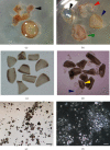
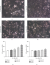
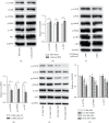
Results: The retina would curl inwardly during the growth medium incubation period, releasing RPE sheets in the medium. Compared with low density group (5,000 cells/cm2), RPE cells plated at high density (15,000 cells/cm2) can maintain RPE morphology for a more extended period. Meanwhile, plating RPE cells at low density significantly reduced the expression of RPE cell type-specific genes (RPE65, CRALBP, and bestrophin) and increased the expression of EMT-related genes (N-cadherin, fibronectin, and α-SMA), in comparison with the samples from the high density group. The petri dish culture condition reduced cell adhesion and thus inhibited RPE cell proliferation. As compared with other culture conditions, RPE cells in the petri dish-N2B27 condition could maintain RPE phenotype with increased expression of RPE-specific genes and decreased expression of EMT-related genes. The AKT/mTOR pathway was also decreased in petri dish-N2B27 condition.
Conclusion: The current study provided an alternative method for easy isolation of RPE cells with high yield and maintenance of its epithelial morphology in the petri dish-N2B27 condition.
Copyright © 2020 Hui Lou et al.
Conflict of interest statement
The authors declare that they have no conflicts of interest.
Figures


Similar articles
-
A cell culture condition that induces the mesenchymal-epithelial transition of dedifferentiated porcine retinal pigment epithelial cells.Exp Eye Res. 2018 Dec;177:160-172. doi: 10.1016/j.exer.2018.08.005. Epub 2018 Aug 8. Exp Eye Res. 2018. PMID: 30096326
-
Artefacts in 1H NMR-based metabolomic studies on cell cultures.MAGMA. 2015 Apr;28(2):161-71. doi: 10.1007/s10334-014-0458-z. Epub 2014 Aug 10. MAGMA. 2015. PMID: 25108704
-
Comparison of two different media for maturation rate of neural progenitor cells to neuronal and glial cells emphasizing on expression of neurotrophins and their respective receptors.Mol Biol Rep. 2018 Dec;45(6):2377-2391. doi: 10.1007/s11033-018-4404-4. Epub 2018 Oct 10. Mol Biol Rep. 2018. PMID: 30306506
-
Numerical modeling and dosimetry of the 35 mm Petri dish under 46 GHz millimeter wave exposure.Bioelectromagnetics. 2005 Sep;26(6):481-8. doi: 10.1002/bem.20121. Bioelectromagnetics. 2005. PMID: 15931681
-
Evaluation of Petri dish sampling for assessment of cat allergen in airborne dust.Allergy. 2002 Feb;57(2):164-8. doi: 10.1034/j.1398-9995.2002.1s3297.x. Allergy. 2002. PMID: 11929422
Cited by
-
Blastocyst-like Structures in the Peripheral Retina of Young Adult Beagles.Int J Mol Sci. 2024 May 30;25(11):6045. doi: 10.3390/ijms25116045. Int J Mol Sci. 2024. PMID: 38892233 Free PMC article.
-
Dihydroartemisinin Inhibits TGF-β-Induced Fibrosis in Human Tenon Fibroblasts via Inducing Autophagy.Drug Des Devel Ther. 2021 Mar 3;15:973-981. doi: 10.2147/DDDT.S280322. eCollection 2021. Drug Des Devel Ther. 2021. PMID: 33688170 Free PMC article.
-
Inhibition of EGFR attenuates EGF-induced activation of retinal pigment epithelium cell via EGFR/AKT signaling pathway.Int J Ophthalmol. 2024 Jun 18;17(6):1018-1027. doi: 10.18240/ijo.2024.06.05. eCollection 2024. Int J Ophthalmol. 2024. PMID: 38895677 Free PMC article.
References
LinkOut - more resources
Full Text Sources
Molecular Biology Databases
Research Materials
Miscellaneous

