

Transfer Learning for Automated OCTA Detection of Diabetic Retinopathy
- PMID: 32855839
- PMCID: PMC7424949
- DOI: 10.1167/tvst.9.2.35
Transfer Learning for Automated OCTA Detection of Diabetic Retinopathy
Abstract
Purpose: To test the feasibility of using deep learning for optical coherence tomography angiography (OCTA) detection of diabetic retinopathy.
Methods: A deep-learning convolutional neural network (CNN) architecture, VGG16, was employed for this study. A transfer learning process was implemented to retrain the CNN for robust OCTA classification. One dataset, consisting of images of 32 healthy eyes, 75 eyes with diabetic retinopathy (DR), and 24 eyes with diabetes but no DR (NoDR), was used for training and cross-validation. A second dataset consisting of 20 NoDR and 26 DR eyes was used for external validation. To demonstrate the feasibility of using artificial intelligence (AI) screening of DR in clinical environments, the CNN was incorporated into a graphical user interface (GUI) platform.
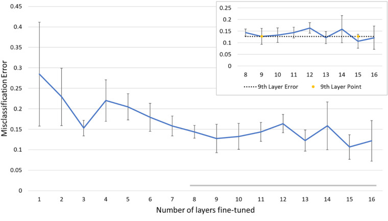
Results: With the last nine layers retrained, the CNN architecture achieved the best performance for automated OCTA classification. The cross-validation accuracy of the retrained classifier for differentiating among healthy, NoDR, and DR eyes was 87.27%, with 83.76% sensitivity and 90.82% specificity. The AUC metrics for binary classification of healthy, NoDR, and DR eyes were 0.97, 0.98, and 0.97, respectively. The GUI platform enabled easy validation of the method for AI screening of DR in a clinical environment.
Conclusions: With a transfer learning process for retraining, a CNN can be used for robust OCTA classification of healthy, NoDR, and DR eyes. The AI-based OCTA classification platform may provide a practical solution to reducing the burden of experienced ophthalmologists with regard to mass screening of DR patients.
Translational relevance: Deep-learning-based OCTA classification can alleviate the need for manual graders and improve DR screening efficiency.
Keywords: artificial intelligence; deep learning; detection; diabetic retinopathy; screening.
Copyright 2020 The Authors.
Conflict of interest statement
Disclosure: D. Le, None; M. Alam, None; C.K. Yao, None; J.I. Lim, None; Y.-T. Hsieh, None; R.V.P. Chan, None; D. Toslak, None; X. Yao, None
Figures


Similar articles
-
A Deep Learning Algorithm for Classifying Diabetic Retinopathy Using Optical Coherence Tomography Angiography.Transl Vis Sci Technol. 2022 Feb 1;11(2):39. doi: 10.1167/tvst.11.2.39. Transl Vis Sci Technol. 2022. PMID: 35703566 Free PMC article.
-
Detection of the Microvascular Changes of Diabetic Retinopathy Progression Using Optical Coherence Tomography Angiography.Transl Vis Sci Technol. 2021 Jun 1;10(7):31. doi: 10.1167/tvst.10.7.31. Transl Vis Sci Technol. 2021. PMID: 34191017 Free PMC article.
-
Statistical Model of Optical Coherence Tomography Angiography Parameters That Correlate With Severity of Diabetic Retinopathy.Invest Ophthalmol Vis Sci. 2018 Aug 1;59(10):4292-4298. doi: 10.1167/iovs.18-24142. Invest Ophthalmol Vis Sci. 2018. PMID: 30167660 Free PMC article.
-
Advancing Diabetic Retinopathy Diagnosis: Leveraging Optical Coherence Tomography Imaging with Convolutional Neural Networks.Rom J Ophthalmol. 2023 Oct-Dec;67(4):398-402. doi: 10.22336/rjo.2023.63. Rom J Ophthalmol. 2023. PMID: 38239418 Free PMC article. Review.
-
Advancing Diabetic Retinopathy Screening: A Systematic Review of Artificial Intelligence and Optical Coherence Tomography Angiography Innovations.Diagnostics (Basel). 2025 Mar 15;15(6):737. doi: 10.3390/diagnostics15060737. Diagnostics (Basel). 2025. PMID: 40150080 Free PMC article. Review.
Cited by
-
An open-source deep learning network AVA-Net for arterial-venous area segmentation in optical coherence tomography angiography.Commun Med (Lond). 2023 Apr 17;3(1):54. doi: 10.1038/s43856-023-00287-9. Commun Med (Lond). 2023. PMID: 37069396 Free PMC article.
-
Multiple instance learning based classification of diabetic retinopathy in weakly-labeled widefield OCTA en face images.Sci Rep. 2023 May 29;13(1):8713. doi: 10.1038/s41598-023-35713-4. Sci Rep. 2023. PMID: 37248309 Free PMC article.
-
A practical guide to optical coherence tomography angiography interpretation.Int J Retina Vitreous. 2020 Nov 13;6(1):55. doi: 10.1186/s40942-020-00262-9. Int J Retina Vitreous. 2020. PMID: 33292740 Free PMC article. Review.
-
Automated Diagnosis of Optical Coherence Tomography Angiography (OCTA) Based on Machine Learning Techniques.Sensors (Basel). 2022 Mar 18;22(6):2342. doi: 10.3390/s22062342. Sensors (Basel). 2022. PMID: 35336513 Free PMC article.
-
Enhanced Deep Learning Model for Classification of Retinal Optical Coherence Tomography Images.Sensors (Basel). 2023 Jun 7;23(12):5393. doi: 10.3390/s23125393. Sensors (Basel). 2023. PMID: 37420558 Free PMC article.
References
-
- National Eye Institute. Eye health data and statistics. Available at: https://nei.nih.gov/eyedata/diabetic. Accessed September 1, 2019.
-
- Nayak J, Bhat PS, Acharya UR, Lim CM, Kagathi M. Automated identification of diabetic retinopathy stages using digital fundus images. J Med Syst. 2008; 32: 107–115. - PubMed
-
- Zahid S, Dolz-Marco R, Freund KB, et al. .. Fractal dimensional analysis of optical coherence tomography angiography in eyes with diabetic retinopathy. Invest Ophthalmol Vis Sci. 2016; 57: 4940–4947. - PubMed
Publication types
MeSH terms
Grants and funding
LinkOut - more resources
Full Text Sources
Medical
