

A Review of Deep Learning for Screening, Diagnosis, and Detection of Glaucoma Progression
- PMID: 32855846
- PMCID: PMC7424906
- DOI: 10.1167/tvst.9.2.42
A Review of Deep Learning for Screening, Diagnosis, and Detection of Glaucoma Progression
Abstract
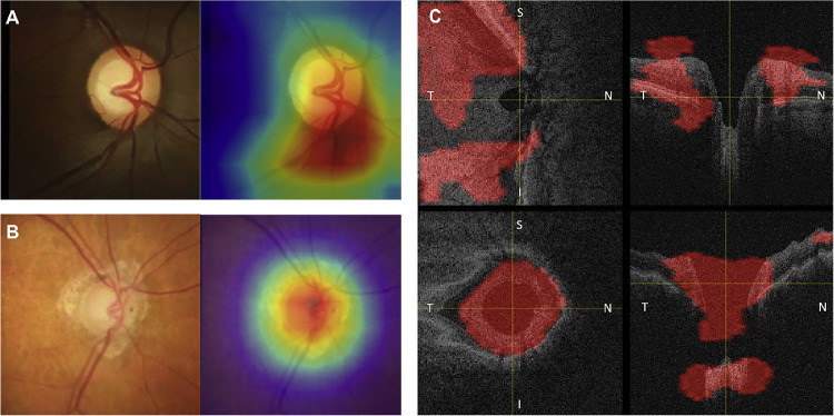
Because of recent advances in computing technology and the availability of large datasets, deep learning has risen to the forefront of artificial intelligence, with performances that often equal, or sometimes even exceed, those of human subjects on a variety of tasks, especially those related to image classification and pattern recognition. As one of the medical fields that is highly dependent on ancillary imaging tests, ophthalmology has been in a prime position to witness the application of deep learning algorithms that can help analyze the vast amount of data coming from those tests. In particular, glaucoma stands as one of the conditions where application of deep learning algorithms could potentially lead to better use of the vast amount of information coming from structural and functional tests evaluating the optic nerve and macula. The purpose of this article is to critically review recent applications of deep learning models in glaucoma, discussing their advantages but also focusing on the challenges inherent to the development of such models for screening, diagnosis and detection of progression. After a brief general overview of deep learning and how it compares to traditional machine learning classifiers, we discuss issues related to the training and validation of deep learning models and how they specifically apply to glaucoma. We then discuss specific scenarios where deep learning has been proposed for use in glaucoma, such as screening with fundus photography, and diagnosis and detection of glaucoma progression with optical coherence tomography and standard automated perimetry.
Translational relevance: Deep learning algorithms have the potential to significantly improve diagnostic capabilities in glaucoma, but their application in clinical practice requires careful validation, with consideration of the target population, the reference standards used to build the models, and potential sources of bias.
Keywords: deep learning; glaucoma; optical coherence tomography; visual fields.
Copyright 2020 The Authors.
Conflict of interest statement
Disclosure: A.C. Thompson, None; A.A. Jammal, None; F.A. Medeiros, Aeri Pharmaceuticals (C); Allergan (C, F), Annexon (C); Biogen (C); Carl Zeiss Meditec (C, F), Galimedix (C); Google Inc. (F); Heidelberg Engineering (F), IDx (C); nGoggle Inc. (P), Novartis (F); Stealth Biotherapeutics (C); Reichert (C, F)
Figures


Similar articles
-
Applications of deep learning in detection of glaucoma: A systematic review.Eur J Ophthalmol. 2021 Jul;31(4):1618-1642. doi: 10.1177/1120672120977346. Epub 2020 Dec 4. Eur J Ophthalmol. 2021. PMID: 33274641
-
[Artificial intelligence and glaucoma: A literature review].J Fr Ophtalmol. 2022 Feb;45(2):216-232. doi: 10.1016/j.jfo.2021.11.002. Epub 2022 Jan 3. J Fr Ophtalmol. 2022. PMID: 34991909 Review. French.
-
Artificial intelligence and complex statistical modeling in glaucoma diagnosis and management.Curr Opin Ophthalmol. 2021 Mar 1;32(2):105-117. doi: 10.1097/ICU.0000000000000741. Curr Opin Ophthalmol. 2021. PMID: 33395111 Review.
-
Interpreting Deep Learning Studies in Glaucoma: Unresolved Challenges.Asia Pac J Ophthalmol (Phila). 2021 May-Jun 01;10(3):261-267. doi: 10.1097/APO.0000000000000395. Asia Pac J Ophthalmol (Phila). 2021. PMID: 34383718 Review.
-
Promising Artificial Intelligence-Machine Learning-Deep Learning Algorithms in Ophthalmology.Asia Pac J Ophthalmol (Phila). 2019 May-Jun;8(3):264-272. doi: 10.22608/APO.2018479. Epub 2019 May 31. Asia Pac J Ophthalmol (Phila). 2019. PMID: 31149787 Review.
Cited by
-
Unmasking biases and navigating pitfalls in the ophthalmic artificial intelligence lifecycle: A narrative review.PLOS Digit Health. 2024 Oct 8;3(10):e0000618. doi: 10.1371/journal.pdig.0000618. eCollection 2024 Oct. PLOS Digit Health. 2024. PMID: 39378192 Free PMC article. Review.
-
Real-Time Risk Score for Glaucoma Mass Screening by Spectral Domain Optical Coherence Tomography: Development and Validation.Transl Vis Sci Technol. 2022 Aug 1;11(8):8. doi: 10.1167/tvst.11.8.8. Transl Vis Sci Technol. 2022. PMID: 35938880 Free PMC article.
-
Machine learning in optical coherence tomography angiography.Exp Biol Med (Maywood). 2021 Oct;246(20):2170-2183. doi: 10.1177/15353702211026581. Epub 2021 Jul 19. Exp Biol Med (Maywood). 2021. PMID: 34279136 Free PMC article. Review.
-
Characteristics of a Large, Labeled Data Set for the Training of Artificial Intelligence for Glaucoma Screening with Fundus Photographs.Ophthalmol Sci. 2023 Mar 17;3(3):100300. doi: 10.1016/j.xops.2023.100300. eCollection 2023 Sep. Ophthalmol Sci. 2023. PMID: 37113471 Free PMC article.
-
Meibography Phenotyping and Classification From Unsupervised Discriminative Feature Learning.Transl Vis Sci Technol. 2021 Feb 5;10(2):4. doi: 10.1167/tvst.10.2.4. Transl Vis Sci Technol. 2021. PMID: 34003889 Free PMC article.
References
-
- Tham YC, Li X, Wong TY, Quigley HA, Aung T, Cheng CY. Global prevalence of glaucoma and projections of glaucoma burden through 2040: a systematic review and meta-analysis. Ophthalmology. 2014; 121: 2081–2090. - PubMed
-
- Hennis A, Wu SY, Nemesure B, Honkanen R, Leske MC, Barbados Eye Studies G. Awareness of incident open-angle glaucoma in a population study: the Barbados Eye Studies. Ophthalmology. 2007; 114: 1816–1821. - PubMed
-
- Harwerth RS, Carter-Dawson L, Smith Barnes ELG III, Holt WF, Crawford MLJ. Neural Losses Correlated with Visual Losses in Clinical Perimetry. Invest Ophthalmol Vis Sci. 2004; 45: 3152–3160. - PubMed
Publication types
MeSH terms
Grants and funding
LinkOut - more resources
Full Text Sources
Medical
Research Materials
