

A Reflectivity Measure to Quantify Bruch's Membrane Calcification in Patients with Pseudoxanthoma Elasticum Using Optical Coherence Tomography
- PMID: 32855880
- PMCID: PMC7422762
- DOI: 10.1167/tvst.9.8.34
A Reflectivity Measure to Quantify Bruch's Membrane Calcification in Patients with Pseudoxanthoma Elasticum Using Optical Coherence Tomography
Abstract
Purpose: Progressive calcification of Bruch's membrane (BM) causes considerable visual morbidity in patients with pseudoxanthoma elasticum (PXE). Since calcification is hyperreflective on optical coherence tomography (OCT), our aim was to measure BM calcification with OCT imaging.
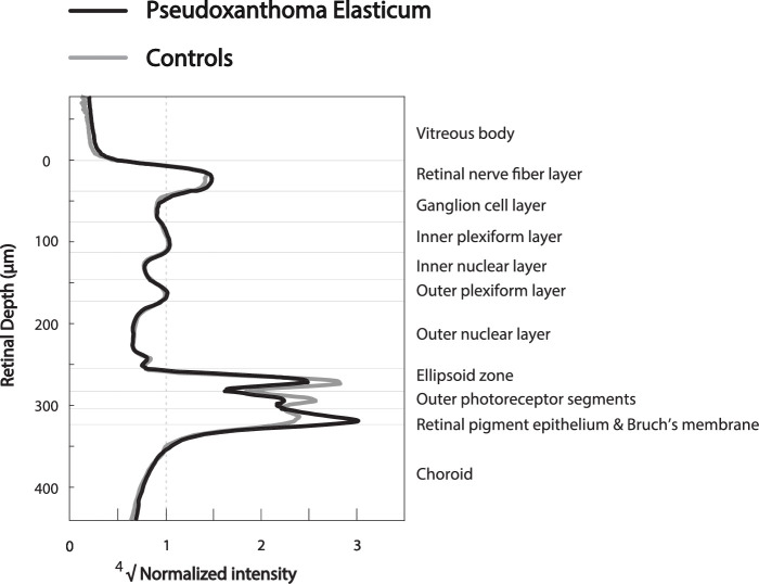
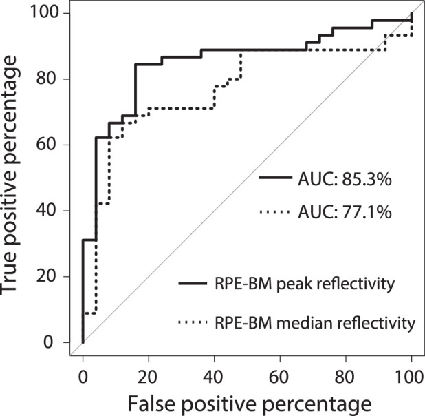
Methods: Case-control study with 45 patients with PXE under 40 years (range, 11-39) and 25 controls (range, 14-39). Spectralis HRA-OCT imaging consisted of seven macular B-scans with 250-µm spacing. Retinal segmentation was performed with the IOWA Reference Algorithms. MATLAB was used to extract and average z-axis reflectivity profiles. Layer reflectivities were normalized to the ganglion cell and inner plexiform layers. Both median and peak layer reflectivities were compared between patients with PXE and controls. The discriminative value of the retinal pigment epithelium (RPE)-BM peak reflectivity was analyzed using receiver operating characteristic analysis.
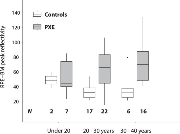
Results: The reflectivity profile of patients with PXE differed from controls in the outer retinal layers. The normalized median RPE-BM reflectivity was 41.1 (interquartile range [IQR], 26.3-51.9) in patients with PXE, compared with 22.5 (IQR, 19.3-29.5) in controls (P = 2.09 × 10⁻³). The normalized RPE-BM peak reflectivity was higher in patients with PXE (67.5; IQR, 42.1-84.2) than in controls (32.7; IQR, 25.7-38.9; P = 2.43 × 10⁻⁵) and had a high discriminative value with an area under the curve of 0.85 (95% confidence interval, 0.76-0.95). In patients with PXE under 40 years, increasing age did not have a statistically significant effect on the RPE-BM peak reflectivity (patients under 20 years: 44.2 [IQR, 40.5-74.6]; 20-30 years: 66.0 [IQR, 45.1-83.8]; 30-40 years: 70.8 [IQR, 49.0-88.0], P = 0.47).
Conclusions: BM calcification can be measured as increased RPE-BM reflectivity in young patients with PXE and has a high discriminative value.
Translational relevance: In patients with PXE, the OCT reflectivity of Bruch's membrane may be the first biomarker for Bruch's membrane calcification and a valuable ophthalmologic endpoint in clinical trials.
Keywords: Bruch's membrane; calcification; pseudoxanthoma elasticum; quantification; reflectivity.
Copyright 2020 The Authors.
Conflict of interest statement
Disclosure: S. Risseeuw, None; E. Bennink, None; M.G. Poirot, None; P.A. de Jong, None; W. Spiering, None; S.M. Imhof, None; R. van Leeuwen, None; J. Ossewaarde-van Norel, None
Figures
References
-
- Bergen AA, Plomp AS, Schuurman EJ, et al. .. Mutations in ABCC6 cause pseudoxanthoma elasticum. Nat Genet. 2000; 25: 228–231. - PubMed
-
- Kranenburg G, Baas AF, de Jong PA, Asselbergs FW, Visseren FLJ, Spiering W. The prevalence of pseudoxanthoma elasticum: revised estimations based on genotyping in a high vascular risk cohort. Eur J Med Genet. 2019; 62: 90–92. - PubMed
-
- Risseeuw S, Ossewaarde-van Norel J, Klaver CCW, Colijn JM, Imhof SM, van Leeuwen R. Visual acuity in pseudoxanthoma elasticum. Retina. 2019; 39: 1580–1587. - PubMed
-
- Kranenburg G, de Jong PA, Bartstra JW, et al. .. Etidronate for prevention of ectopic mineralization in patients with pseudoxanthoma elasticum. J Am Coll Cardiol. 2018; 71: 1117–1126. - PubMed
Publication types
MeSH terms
LinkOut - more resources
Full Text Sources
