

Forehead Flap Ballooning for Scar Revision
- PMID: 32855937
- PMCID: PMC7433955
- DOI: 10.4103/ams.ams_266_19
Forehead Flap Ballooning for Scar Revision
Abstract
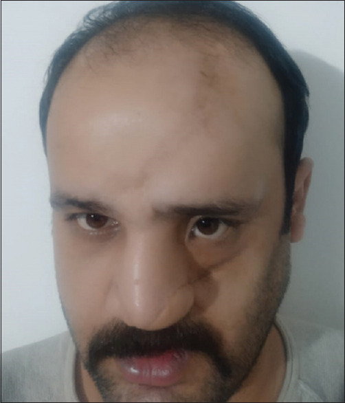
Nowadays, cutaneous expansion is used progressively in reconstructive surgery for treating the variety of problems such as burns alopecia scar revision in children and adults. With the use of tissue expansion technique, the reconstruction of many acquired and congenital defects has been made possible. Tissue expanders are principally based on the mechanical and the biological creep in which mechanical creep is the morphological changes occurring in cellular level in response to applied stress and biological creep is the resultant expansion of skin surface. There is an excellent closure of extensive soft tissue defects without additional scars in donor area with tissue expanders as compared with other methods of plastic surgery the case report highlights the excellent results of tissue expander in an esthetically compromised patient due to hypotrophic scar on the forehead. This is a novel technique as it was performed under local anesthesia without the use of any sedation in a regular clinical setup.
Keywords: Hypotrophic scar revision; reconstruction; tissue expander.
Copyright: © 2020 Annals of Maxillofacial Surgery.
Conflict of interest statement
There are no conflicts of interest.
Figures


Similar articles
-
Optimization of the use of skin expanders.Skin Res Technol. 2014 Nov;20(4):463-72. doi: 10.1111/srt.12141. Epub 2014 Feb 17. Skin Res Technol. 2014. PMID: 24527999
-
The feasibility of tissue expansion in reconstruction of congenital and aquired deformities of pediatric patients.Int J Burns Trauma. 2013 Jul 8;3(3):144-50. Print 2013. Int J Burns Trauma. 2013. PMID: 23875120 Free PMC article.
-
A new technique of scarless expanded forehead flap for reconstructive surgery.Plast Reconstr Surg. 2000 Sep;106(4):777-85. doi: 10.1097/00006534-200009040-00004. Plast Reconstr Surg. 2000. PMID: 11007388
-
Reconstructive surgery of extensive face and neck burn scars using tissue expanders.World J Plast Surg. 2015 Jan;4(1):40-9. World J Plast Surg. 2015. PMID: 25606476 Free PMC article.
-
Surgery for scar revision and reduction: from primary closure to flap surgery.Burns Trauma. 2019 Mar 1;7:7. doi: 10.1186/s41038-019-0144-5. eCollection 2019. Burns Trauma. 2019. PMID: 30891462 Free PMC article. Review.
References
-
- Motamed S, Niazi F, Atarian S, Motamed A. Post-burn head and neck reconstruction using tissue expanders. Burns. 2008;34:878–84. - PubMed
-
- van Rappard JH, Sonneveld GJ, Boughouts JM. Geometric planning and the shape of the expander. In: van Rappard JH, editor. Tissue Expansion in Facial Plastic Surgery. Vol. 5. New York: Thieme; 1988. - PubMed
-
- Awad M. The effect of tissue expanders on the growing craniofacial skeleton. Indian J Plast Surg. 2006;1:22.
-
- Sharobaro VI, Moroz VY, Starkov YG, Strekalovsky VP. First experience of endoscopic implantation of tissue expanders in plastic and reconstructive surgery. Surg Endosc. 2004;18:513–7. - PubMed
LinkOut - more resources
Full Text Sources