. 2020 Sep;9(17):e015307.
doi: 10.1161/JAHA.119.015307. Epub 2020 Aug 28.

Deletion of Microfibrillar-Associated Protein 4 Attenuates Left Ventricular Remodeling and Dysfunction in Heart Failure

Affiliations

Deletion of Microfibrillar-Associated Protein 4 Attenuates Left Ventricular Remodeling and Dysfunction in Heart Failure

Hui-Bo Wang et al. J Am Heart Assoc. 2020 Sep.

Abstract

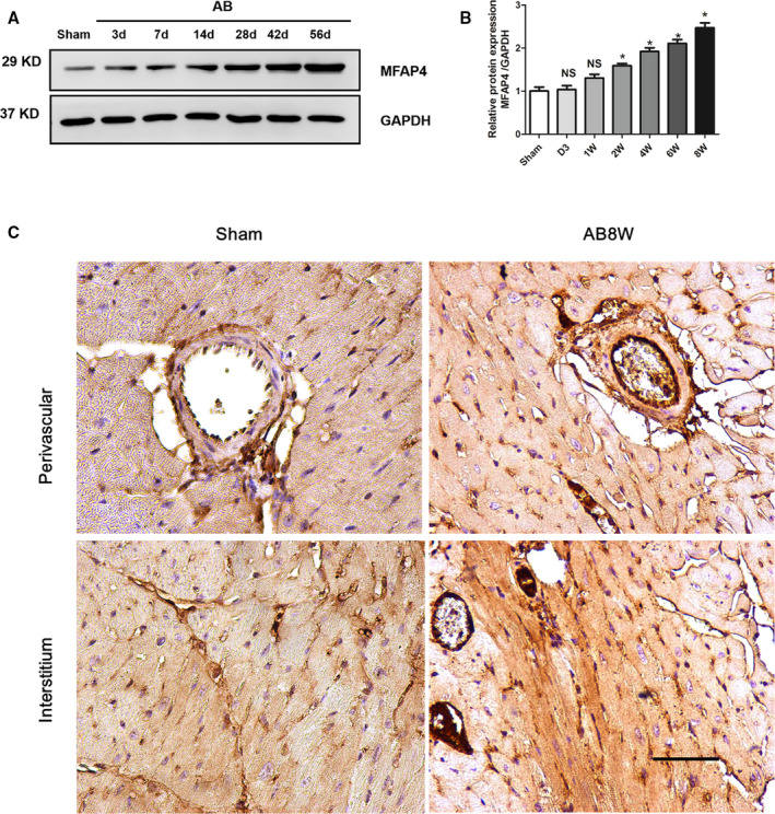
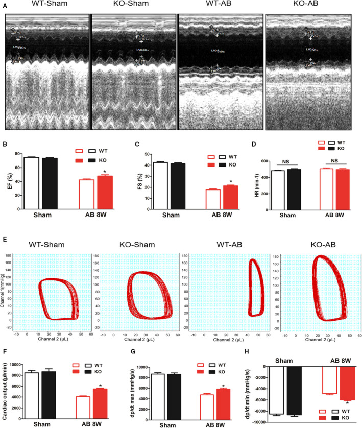
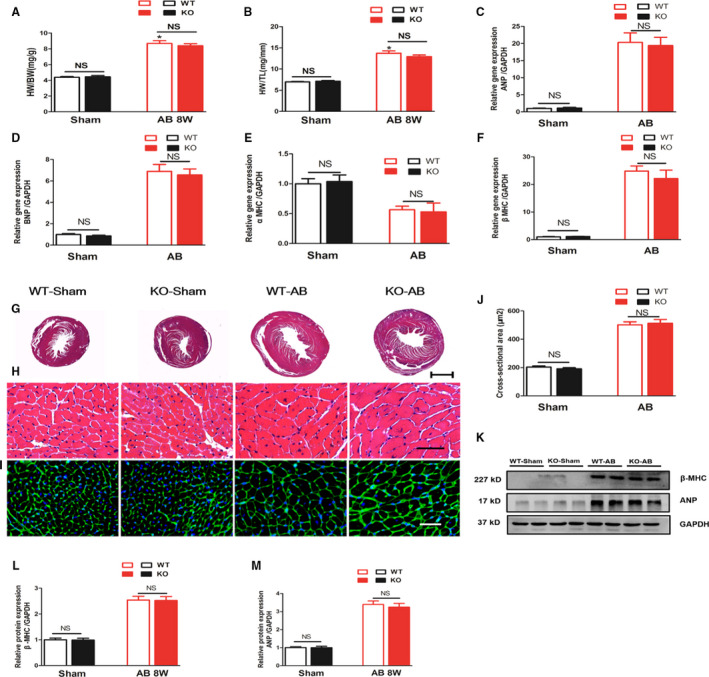
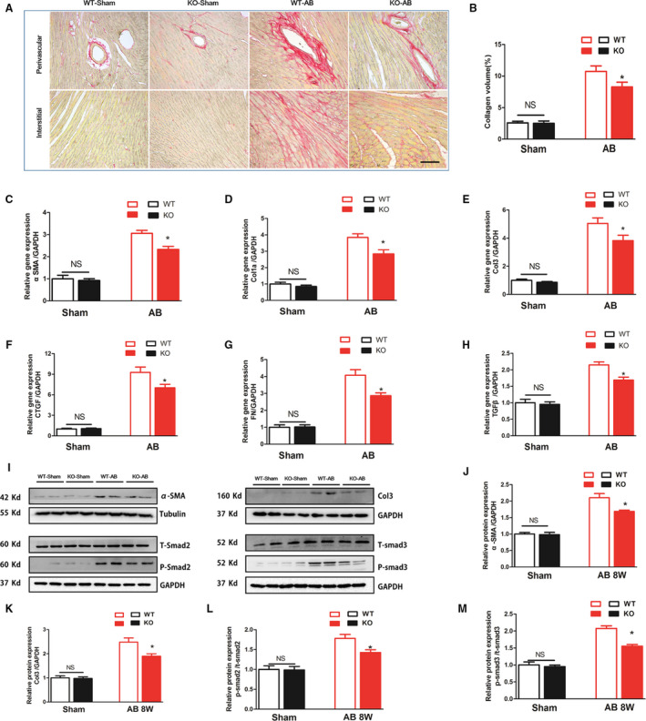
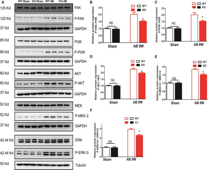
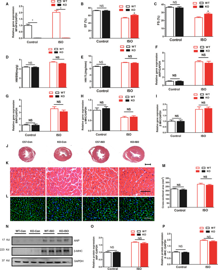
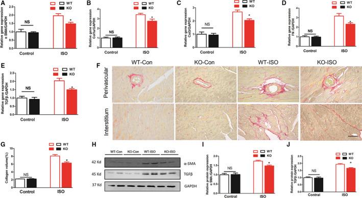
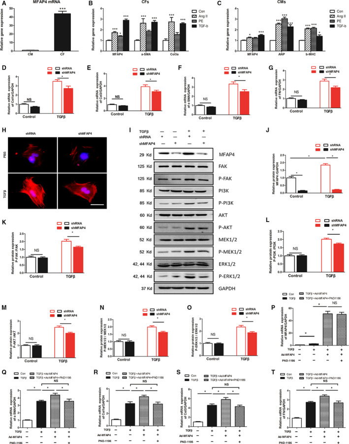
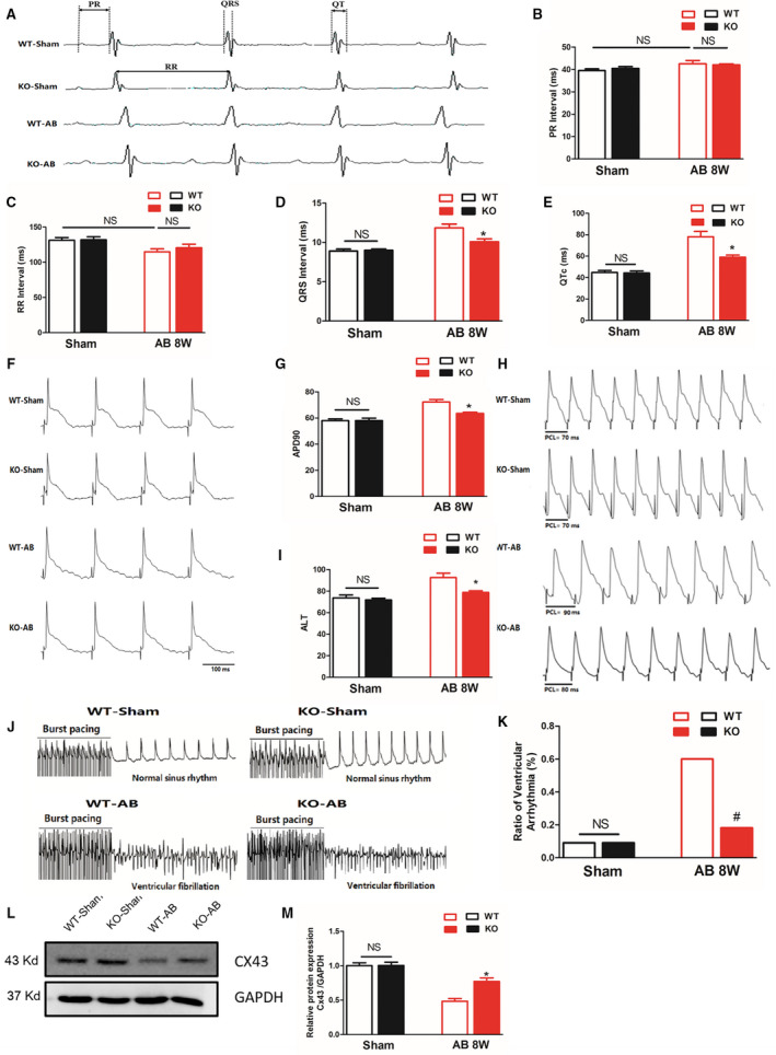
Background Cardiac remodeling predisposes individuals to heart failure if the burden is not solved, and heart failure is a growing cause of morbidity and mortality worldwide. The cardiac extracellular matrix not only provides structural support, but also is a core aspect of the myocardial response to various biomechanical stresses and heart failure. MFAP4 (microfibrillar-associated protein 4) is an integrin ligand located in the extracellular matrix, whose biological functions in the heart remain poorly understood. In the current study we aimed to test the role of MFAP4 in cardiac remodeling. Methods and Results MFAP4-deficient (MFAP4-/-) and wild-type mice were subjected to aortic banding surgery and isoproterenol to establish models of cardiac remodeling. We also evaluated the functional effects of MFAP4 on cardiac hypertrophy, fibrosis, and cardiac electrical remodeling. The expression of MFAP4 was increased in the animal cardiac remodeling models induced by pressure overload and isoproterenol. After challenge of 8 weeks of aortic banding or 2 weeks of intraperitoneal isoproterenol, MFAP4-/- mice exhibited lower levels of cardiac fibrosis and fewer ventricular arrhythmias than wild-type mice. However, there was no significant effect on cardiomyocyte hypertrophy. In addition, there was no significant difference in cardiac fibrosis severity, hypertrophy, or ventricular arrhythmia incidence between wild-type-sham and knockout-sham mice. Conclusions These findings are the first to demonstrate that MFAP4 deficiency inhibits cardiac fibrosis and ventricular arrhythmias after challenge with 8 weeks of aortic banding or 2 weeks of intraperitoneal isoproterenol but does not significantly affect the hypertrophy response. In addition, MFAP4 deficiency had no significant effect on cardiac fibrosis, hypertrophy, or ventricular arrhythmia in the sham group in this study.

Keywords: cardiac remodeling; extracellular matrix proteins; heart failure; microfibrillar-associated protein 4; pressure overload.


Conflict of interest statement

None.

Figures

Figure 1
Figure 1. MFAP4 is overexpressed in response to pressure overload.
A and B, Representative western blot of MFAP4 (microfibrillar‐associated protein 4) in heart tissues after aortic banding at the indicated time points (3, 7, 14, 28, 42, and 56 days) (n=5). C, Immunohistochemical detection of MFAP4 expression in the sham group and the 8‐week aortic banding group. Scale bar: 50 μm (n=3). AB indicates aortic banding; AB 8W, 8‐week aortic banding group; and MFAP4, microfibrillar‐associated protein 4. *P<0.05 compared with Sham group.
Figure 2
Figure 2. MFAP4 deficiency alleviates cardiac dysfunction after pressure overload.
A, Representative echocardiographic images for each group. B through D, Statistical results for the ejection fraction, left ventricular fractional shortening, and heart rate (n=15 for the wild‐type‐sham and knockout‐sham groups; n=12 for the wild‐type‐aortic banding [AB] and knockout‐AB groups). E, Representative pressure‐volume loop images for each group. F through H, Statistical results for the cardiac output, maximal rate of pressure development (dP/dtmax), and maximal rate of pressure decay (dP/dtmin) (n=12 for the WT‐sham and knockout‐sham groups n=11 for the WT‐AB and knockout‐AB groups). AB indicates aortic banding; CO, cardiac output; dp/dtmax, maximal rate of left ventricle pressure development; dp/dtmin, minimal rate of left ventricle pressure development; EF, ejection fraction; FS, fractional shortening; and HR, heart rate. *P<0.05 represents the comparison between WT‐AB and KO‐AB group.
Figure 3
Figure 3. MFAP4 deficiency does not improve aortic banding‐induced cardiac hypertrophy.
A and B, heart weight/body weight and heart weight/tibia length ratios of wild‐type (WT) and MFAP4‐knockout mice (KO) after sham or aortic banding (AB) operation (n=15 for the WT‐sham and KO‐sham groups; n=16 for the WT‐AB and KO‐AB groups). C through F, The effects of AB on the mRNA expression of ANP, BNP, α‐MHC, and β‐MHC were determined by reverse transcription‐polymerase chain reaction analysis (n=6). G and H, Representative hematoxylin and eosin‐stained heart sections of WT or MFAP4‐KO mice after sham or AB operation. G, 10× (scale bar: 400 μm), (H) 400× (scale bar: 50 μm). n=6. I, Representative wheat germ agglutinin‐fluorescein isothiocyanate isomer staining of sham and AB mice at 8 weeks post‐surgery, Scale bar: 50 μm. J, Quantification of the CSA in each group (n=6). K through M, Representative blots and quantitative results for ANP and β‐MHC protein expression in myocardium in each group (n=6). AB indicates aortic banding; AB 8W, 8‐week aortic banding group; ANP indicates atrial natriuretic peptide; BNP, B‐type natriuretic peptide; HW/BW, heart weight/body weight; HW/TL, heart weight/tibia length; KO, knockout mice; NS, no statistically difference; WGA, wheat germ agglutinin; and β‐MHC, β‐myosin heavy polypeptide. *P<0.05 compared with the corresponding sham group.
Figure 4
Figure 4. MFAP4 deficiency attenuates aortic banding‐induced cardiac fibrosis.
A, Representative image of the heart with Picrosirius staining. scale bar: 50 μm. B, Quantification of the total collagen volume in the indicated group (n=6). C through H, PCR analysis of fibrotic markers (αSMA, collagen Iα, collagen III, CTGF, fibronectin, TGF‐β) (n=6). I through M, Representative blots and quantitative results for αSMA, collagen III and P‐Smad2/T‐Smad2, and P‐Smad3/T‐Smad3 protein expression in the myocardium in each group (n=6). AB indicates aortic banding; Col1α, collagen Iα; Col3, collagen type 3; CTGF, connective tissue growth factor; KO, knockout mice; MFAP4, microfibrillar‐associated protein 4; PSR, Picrosirius red; TGF‐β, transforming growth factor‐β; WT, wild‐type; and αSMA, alpha‐smooth muscle actin. *P<0.05 compared with the corresponding wild‐type‐aortic banding group.
Figure 5
Figure 5. MFAP4 deficiency significantly inhibits FAK and downstream signaling.
A through F, Representative blots and quantitative results for P‐FAK/T‐FAK, P‐PI3K/T‐PI3K, P‐AKT/T‐AKT, P‐MEK1/2/ T‐MEK1/2, P‐ERK1/2/T‐ERK1/2 protein expression in the myocardium in each group (n=6). *P<0.05 compared with the corresponding WT‐AB group. AB indicates aortic banding; ERK, extracellular signal regulated kinase; FAK, focal adhesion kinase; KO, knockout mice; MEK, mitogen‐activated protein kinase (MAPK)/ERK kinase; PI3K, phosphatidylinositol 3 kinase; and WT, wild‐type.
Figure 6
Figure 6. MFAP4 deficiency does not improve isoproterenol‐induced cardiac hypertrophy.
A, Polymerase chain reaction analysis of MFAP4 mRNA expression in the myocardium in each group (n=6). B and C, Statistical results for the ejection fraction and fractional shortening in the indicated group (n=10 for the WT‐Control and knockout mice (KO)‐Control groups; n=13 for the WT‐isoproterenol group; n=12 for the KO‐isoproterenol group). D and E, HW/BW and HW/tibia length ratios of WT and MFAP4‐KO mice after vehicle (control) or isoproterenol IP injection (n=12 for the WT‐Control and KO‐Con groups; n=23 for the WT‐isoproterenol group; n=13 for the KO‐isoproterenol group). F through I, The effects of isoproterenol on the mRNA expression of ANP, BNP, α‐MHC, and β‐MHC were determined by reverse transcription‐polymerase chain reaction analysis (n=6). J through L, Hematoxylin and eosin staining and wheat germ agglutinin‐fluorescein isothiocyanate isomer staining of the control or isoproterenol mice. J, 10× (scale bar: 400 μm), (K) 400× (scale bar: 50 μm). M, Quantification of the cross‐sectional areas in each group (n=6). N through P, Representative blots and quantitative results for ANP and β‐MHC protein expression in the myocardium in each group (n=6). ANP indicates atrial natriuretic peptide; HW/BW, heart weight/body weight; HW/TL, heart weight/tibia length; KO, knockout mice; NS, no statistically difference; WGA, wheat germ agglutinin; WT, wild‐type; and β‐MHC, β‐myosin heavy polypeptide. *P<0.05 compared with the corresponding control group.
Figure 7
Figure 7. MFAP4 deficiency attenuated isoprotenerol‐induces cardiac fibrosis.
A through E, Polymerase chain reaction analysis of fibrotic markers (αSMA, collagen Iα, collagen III, fibronectin, transforming growth factor‐β) in the myocardium in each group (n=6). F, Representative image of the heart with Picrosirius staining. G, Quantification of the total collagen volume in the indicated group (n=6). H and I, Representative blots and quantitative results for ANP and β‐MHC protein expression in the myocardium in each group (n=6). Col1α indicates collagen Iα; CTGF, connective tissue growth factor; KO, knockout mice; MFAP4, microfibrillar‐associated protein 4; PSR, Picrosirius red; TGF‐β, transforming growth factor‐β; WT, wild‐type; and αSMA, alpha‐smooth muscle actin. *P<0.05 compared with the corresponding wild‐type‐isoproterenol group.
Figure 8
Figure 8. Role and mechanism of MFAP4 in cardiac fibroblasts
A, Polymerase chain reaction analysis of mRNA level of MFAP4 (microfibrillar‐associated protein 4) between cardiomyocytes and cardiac fibroblasts (n=6). The data were compared by unpaired Student t‐test. B and C, Polymerase chain reaction analysis of the mRNA expression level of MFAP4 and markers of, hypertrophic and fibrosis markers when in cardiomyocytes and cardiac fibroblasts were stimulated by angiotensin II (Ang II), phenylephrine, and or TGF‐β, respectively (n=6). (D through G) PCR analysis of the mRNA expression levels of αSMA, Col1α, Col3, and fibronectin (n=6). H, Immunofluorescence staining of αSMA (n=6). I through O, Representative blots and quantitative results for P‐FAK/T‐FAK, P‐PI3K/T‐PI3K, P‐AKT/T‐AKT, P‐MEK1/2/T‐MEK1/2, P‐ERK1/2/T‐ERK1/2 protein expression in cardiac fibroblasts each group (n=4). P through T, Polymerase chain reaction analysis of the mRNA expression levels of MFAP4 and fibrosis markers in cardiac fibroblasts treated with Ad‐MFAP4 and an inhibitor of FAK (PND‐1186) (n=6). Ang II indicates angiotensin II; CF, cardiac fibroblasts; CMs, Cardiomyocyte; Col1α, collagen Iα; Col3, collagen 3; CTGF, connective tissue growth factor; ERK, extracellular signal regulated kinase; FAK, focal adhesion kinase; ; MEK, mitogen‐activated protein kinase (MAPK)/ERK kinase; MFAP4, microfibrillar‐associated protein 4; PI3K, phosphatidylinositol 3kinase; PSR, Picrosirius red; TGF‐β, transforming growth factor‐β; and αSMA, alpha‐smooth muscle actin. *P<0.05; **P<0.01; *P<0.001.
Figure 9
Figure 9. MFAP4 deficiency alleviates aortic banding‐induced ventricular arrhythmia.
A, Representative ECG recordings and a comparison of ECG parameters. B through E, Statistical analysis of the PR, RR, QRS, and QTc intervals in MFAP4 (microfibrillar‐associated protein 4)‐knockout (KO) and wild‐type (WT) mice 8 weeks after the sham operation or aortic banding (AB) surgery (n=8 for the WT‐sham and KO‐sham groups; n=13 for the WT‐AB group; n=7 for the knockout [KO]‐AB group). F and G, Representative action potential figures and statistical analysis of the APD90 values in MFAP4‐KO and WT mice 8 weeks after the sham operation or AB surgery (n=8). H and I, Representative electric alternans figures and statistical analysis of the ALT thresholds in MFAP4‐KO and WT mice 8 weeks after the sham operation or AB surgery (n=8). J and K, Representative arrhythmia induced by burst‐pacing stimulation and statistical analysis of MFAP4‐KO and WT mice 8 weeks after the sham operation or AB surgery (n=11 for the WT‐sham, KO‐sham, and KO‐AB groups; n=10 for the WT‐AB group). L and M, Representative western blots, and statistical analysis of electroconduction‐related protein (Connexin 43) levels in MFAP4‐KO and WT mouse heart tissues (n=4). AB indicate aortic banding; AB 8W, 8‐week aortic banding group; ALT, action potential duration alternans; APD90, 90% action potential duration; Cx43, Connexin 43; HW/BW, heart weight/body weight; KO, knockout mice; MFAP4, microfibrillar‐associated protein 4; and WT, wild‐type. *P<0.05 represents the comparison between WT‐AB and KO‐AB group.

Similar articles

Cited by

References

    1. Schuttler D, Clauss S, Weckbach LT, Brunner S. Molecular mechanisms of cardiac remodeling and regeneration in physical exercise. Cells. 2019;8:1128. - PMC - PubMed
    1. Cohn JN, Ferrari R, Sharpe N. Cardiac remodeling–concepts and clinical implications: a consensus paper from an international forum on cardiac remodeling. Behalf of an international forum on cardiac remodeling. J Am Coll Cardiol. 2000;35:569–582. - PubMed
    1. Misra A, Mann DL. Treatment of heart failure beyond practice guidelines. Role of cardiac remodeling. Circ J. 2008;72(suppl A):A1–A7. - PubMed
    1. Aimo A, Gaggin HK, Barison A, Emdin M, Januzzi JL Jr. Imaging, biomarker, and clinical predictors of cardiac remodeling in heart failure with reduced ejection fraction. JACC Heart Fail. 2019;7:782–794. - PubMed
    1. Wang HB, Duan MX, Xu M, Huang SH, Yang J, Yang J, Liu LB, Huang R, Wan CX, Ma ZG, et al. Cordycepin ameliorates cardiac hypertrophy via activating the ampkalpha pathway. J Cell Mol Med. 2019;23:5715–5727. - PMC - PubMed

Publication types

MeSH terms

LinkOut - more resources