

Oral pigmented lesions: a retrospective analysis from Brazil
- PMID: 32856618
- PMCID: PMC8141314
- DOI: 10.4317/medoral.24168
Oral pigmented lesions: a retrospective analysis from Brazil
Abstract
Background: Pigmented lesions are uncommon in the oral mucosa, and studies investigating the incidence and types of these lesions are desired to improve the diagnostic knowledge of clinicians. The aim of this study was to analyze the distribution of oral pigmented lesions in a Brazilian population.
Material and methods: A retrospective descriptive cross-sectional study was performed. Oral pigmented lesions were retrieved from the files of two oral and maxillofacial pathology services from Brazil over a 45-year period (1974-2019). The clinical data and the diagnoses of each case were retrieved and included in a Microsoft Excel® database.
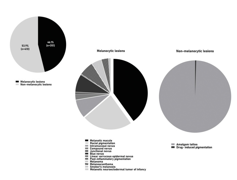
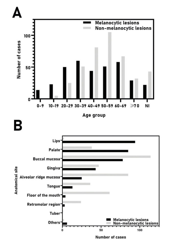
Results: From 77.074 lesions diagnosed in this period, 761 (0.99%) represented pigmented lesions of the oral mucosa, including 351 (46.1%) melanocytic and 410 (53.9%) non-melanocytic lesions, with a higher incidence in females (73.2%) between the fourth and seventh decades of life. Amalgam tattoo (53.6%) represented the most common lesion, followed by melanotic macule (18.3%) and racial pigmentation (10.8%). Other pigmented lesions included nevus (9.9%), post-inflammatory pigmentation (3%), melanoma (2.1%), melanoacanthoma (1.4%), smoker's melanosis (0.4%), drug-induced pigmentation (0.3%), and melanotic neuroectodermal tumor of infancy (0.1%). The buccal mucosa was the most commonly affected site (25.2%), followed by the alveolar ridge (14.5%), and gingiva (11.8%).
Conclusions: The current findings were similar to previous studies with minor differences due methodology and characteristics of the services from where lesions were retrieved. The knowledge of these data may contribute to a better understanding of oral pigmented lesions and assist clinicians to better recognize and manage them.
Conflict of interest statement
Conflicts of interest The authors declare that they have no conflict of interest.
Figures
References
-
- Tavares TS, Meirelles DP, de Aguiar MCF, Caldeira PC. Pigmented lesions of the oral mucosa: A cross-sectional study of 458 histopathological specimens. Oral Dis. 2018;24:1484–91. - PubMed
-
- Hassona Y, Sawair F, Al-Karadsheh O, Scully C. Prevalence and clinical features of pigmented oral lesions. Int J Dermatol. 2016;55:1005–13. - PubMed
-
- Buchner A, Merrell PW, Carpenter WM. Relative frequency of solitary melanocytic lesions of the oral mucosa. J Oral Pathol Med. 2004;33:550–7. - PubMed
-
- Müller S. Melanin-associated pigmented lesions of the oral mucosa: presentation, differential diagnosis, and treatment. Dermatol Ther. 2010;23:220–9. - PubMed
MeSH terms
LinkOut - more resources
Full Text Sources
Medical
