

The promise of placental extracellular vesicles: models and challenges for diagnosing placental dysfunction in utero†
- PMID: 32856695
- PMCID: PMC7786267
- DOI: 10.1093/biolre/ioaa152
The promise of placental extracellular vesicles: models and challenges for diagnosing placental dysfunction in utero†
Abstract
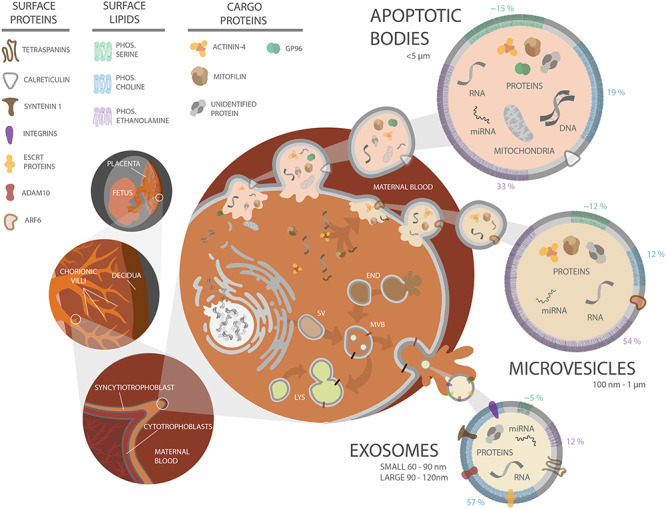
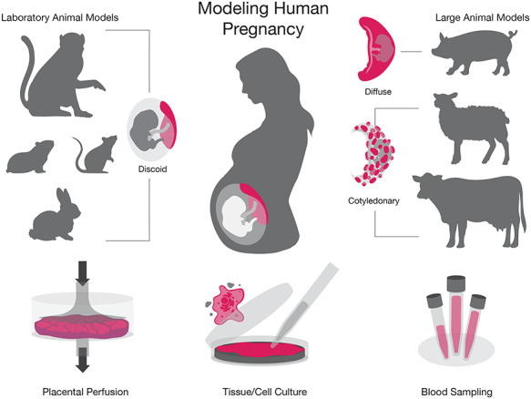
Monitoring the health of a pregnancy is of utmost importance to both the fetus and the mother. The diagnosis of pregnancy complications typically occurs after the manifestation of symptoms, and limited preventative measures or effective treatments are available. Traditionally, pregnancy health is evaluated by analyzing maternal serum hormone levels, genetic testing, ultrasonographic imaging, and monitoring maternal symptoms. However, researchers have reported a difference in extracellular vesicle (EV) quantity and cargo between healthy and at-risk pregnancies. Thus, placental EVs (PEVs) may help to understand normal and aberrant placental development, monitor pregnancy health in terms of developing placental pathologies, and assess the impact of environmental influences, such as infection, on pregnancy. The diagnostic potential of PEVs could allow for earlier detection of pregnancy complications via noninvasive sampling and frequent monitoring. Understanding how PEVs serve as a means of communication with maternal cells and recognizing their potential utility as a readout of placental health have sparked a growing interest in basic and translational research. However, to date, PEV research with animal models lags behind human studies. The strength of animal pregnancy models is that they can be used to assess placental pathologies in conjunction with isolation of PEVs from fluid samples at different time points throughout gestation. Assessing PEV cargo in animals within normal and complicated pregnancies will accelerate the translation of PEV analysis into the clinic for potential use in prognostics. We propose that appropriate animal models of human pregnancy complications must be established in the PEV field.
Keywords: adverse pregnancy outcomes; animal models; exosome; extracellular vesicle; placenta.
© The Author(s) 2020. Published by Oxford University Press on behalf of Society for the Study of Reproduction. All rights reserved. For permissions, please e-mail: journals.permissions@oup.com.
Figures
Similar articles
-
Placenta Extracellular Vesicles: Messengers Connecting Maternal and Fetal Systems.Biomolecules. 2024 Aug 13;14(8):995. doi: 10.3390/biom14080995. Biomolecules. 2024. PMID: 39199382 Free PMC article. Review.
-
Augmented Placental Protein 13 in Placental-Associated Extracellular Vesicles in Term and Preterm Preeclampsia Is Further Elevated by Corticosteroids.Int J Mol Sci. 2023 Jul 27;24(15):12051. doi: 10.3390/ijms241512051. Int J Mol Sci. 2023. PMID: 37569423 Free PMC article.
-
Human placenta releases extracellular vesicles carrying corticotrophin releasing hormone mRNA into the maternal blood.Placenta. 2024 Feb;146:71-78. doi: 10.1016/j.placenta.2024.01.004. Epub 2024 Jan 5. Placenta. 2024. PMID: 38190772
-
Placental exosomes in normal and complicated pregnancy.Am J Obstet Gynecol. 2015 Oct;213(4 Suppl):S173-81. doi: 10.1016/j.ajog.2015.07.001. Am J Obstet Gynecol. 2015. PMID: 26428497 Review.
-
Antiphospholipid antibodies do not cause retargeting of placental extracellular vesicles in the maternal body.Placenta. 2022 Feb;118:66-69. doi: 10.1016/j.placenta.2022.01.008. Epub 2022 Jan 13. Placenta. 2022. PMID: 35042085
Cited by
-
The revolutionary role of placental derivatives in biomedical research.Bioact Mater. 2025 Mar 19;49:456-485. doi: 10.1016/j.bioactmat.2025.03.011. eCollection 2025 Jul. Bioact Mater. 2025. PMID: 40177109 Free PMC article. Review.
-
Clinical Assessment of Fetal Well-Being and Fetal Safety Indicators.J Clin Pharmacol. 2022 Sep;62 Suppl 1(Suppl 1):S67-S78. doi: 10.1002/jcph.2126. J Clin Pharmacol. 2022. PMID: 36106777 Free PMC article. Review.
-
Increased Expression of Tetraspanins, ALIX and HSP-70 in the Placenta Tissue of Women with Chronic Venous Disease during Pregnancy.Int J Med Sci. 2023 Oct 16;20(13):1744-1754. doi: 10.7150/ijms.87830. eCollection 2023. Int J Med Sci. 2023. PMID: 37928882 Free PMC article.
-
Placenta Extracellular Vesicles: Messengers Connecting Maternal and Fetal Systems.Biomolecules. 2024 Aug 13;14(8):995. doi: 10.3390/biom14080995. Biomolecules. 2024. PMID: 39199382 Free PMC article. Review.
-
The ruminant placental trophoblast binucleate cell: an evolutionary breakthrough.Biol Reprod. 2022 Sep 12;107(3):705-716. doi: 10.1093/biolre/ioac107. Biol Reprod. 2022. PMID: 35594454 Free PMC article. Review.
References
-
- Blencowe H, Cousens S, Oestergaard MZ, Chou D, Moller AB, Narwal R, Adler A, Vera Garcia C, Rohde S, Say L, Lawn JE. National, regional, and worldwide estimates of preterm birth rates in the year 2010 with time trends since 1990 for selected countries: a systematic analysis and implications. Lancet 2012; 379:2162–2172. - PubMed
-
- Lawn JE, Kerber K, Enweronu-Laryea C, Cousens S. 3.6 million neonatal deaths--what is progressing and what is not? Semin Perinatol 2010; 34:371–386. - PubMed
-
- Harrison MS, Goldenberg RL. Global burden of prematurity. Semin Fetal Neonatal Med 2016; 21:74–79. - PubMed
-
- Swanson AM, David AL. Animal models of fetal growth restriction: considerations for translational medicine. Placenta 2015; 36:623–630. - PubMed
