WT1 Clone 6F-H2 Cytoplasmic Expression Differentiates Astrocytic Tumors from Astrogliosis and Associates with Tumor Grade, Histopathology, IDH1 Status, Apoptotic and Proliferative Indices: A Tissue Microarray Study
- PMID: 32856872
- PMCID: PMC7771928
- DOI: 10.31557/APJCP.2020.21.8.2403
WT1 Clone 6F-H2 Cytoplasmic Expression Differentiates Astrocytic Tumors from Astrogliosis and Associates with Tumor Grade, Histopathology, IDH1 Status, Apoptotic and Proliferative Indices: A Tissue Microarray Study
Abstract
Objectives: This tissue microarray (TMA) immunohistochemical (IHC) study elucidates the role of Wilms' tumor 1 protein (WT1) in diagnosis and prognostication of astrocytic tumors.
Methods: IHC was applied to 75 astrocytic lesions (18 astrogliosis and 57 astrocytic tumors) using antibodies directed against WT1 clone 6F-H2, isocitrate dehydrogenase 1(IDH1), Bcl2 and Ki67. WT1 IHC staining was evaluated and scored blindly by 2 pathologists. Bcl2 and Ki67 scores and labelling indices were assessed and IDH1 status determined. Statistical analysis was performed using the appropriate methodology.
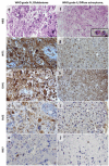
Results: WT1 cytoplasmic expression was detected in 89.5% of astrocytic tumors but not in astrogliosis. Positive WT1 differentiated astrocytic tumors (92.6% accuracy) and grade II diffuse astrocytomas (93.5% accuracy) from astrogliosis with high sensitivity, specificity and positive predictive values (p<0.001). Increased WT1 score significantly associated higher Bcl2 and Ki67 labelling indices, increasing WHO tumor grade and tumor's histopathologic type (p<0.05) showing staining pattern variability by tumor entity and cell type. Glioblastomas, gliosarcomas and subependymal giant cell astrocytomas were the most frequently WT1 expressing tumors with frequent +3 WT1 score. About 21.4% of pilocytic astrocytomas had +3WT1 score in association with increased Bcl2 and Ki67 indices. Low WT1 scores in grade II and III diffuse astrocytomas were linked to the high frequency of IDH1 positivity, and were associated with low Bcl2 and Ki67 labelling indices. In glioblastomas, WT1 significantly associated poor prognostic variables: older age, negative-IDH1 status, high Bcl2 and Ki67 labelling indices (p=0.04, <0.001, =0.001 and.
Keywords: Astrocytic tumors; Astrogliosis; Bcl2/Ki67; IDH1; WT1.
Figures


References
-
- Ambroise MM, Khosla C, Ghosh M, Mallikarjuna VS, Annapurneswari S. The role of immunohistochemistry in predicting behavior of astrocytic tumors. Asian Pac J Cancer Prev. 2010;11:1079–84. - PubMed
-
- Bassam AM, Abdel-Salam LO, Khairy D. WT1 Expression in glial tumors: Its possible role in angiogenesis and prognosis. Academic J Cancer Res. 2014;7:50–8.
-
- Camacho-Urkaray E, Santos-Juanes J, Gutiérrez-Corres FB, et al. Establishing cut-off points with clinical relevance for bcl-2, cyclin D1, p16, p21, p27, p53, Sox11 and WT1 expression in glioblastoma - a short report. Cell Oncol (Dordr) 2018;41:213–21. - PubMed
MeSH terms
Substances
LinkOut - more resources
Full Text Sources
Miscellaneous

