Antitumor Effects of Fucoidan Via Apoptotic and Autophagic Induction on HSC-3 Oral Squamous CellCarcinoma
- PMID: 32856880
- PMCID: PMC7771925
- DOI: 10.31557/APJCP.2020.21.8.2469
Antitumor Effects of Fucoidan Via Apoptotic and Autophagic Induction on HSC-3 Oral Squamous CellCarcinoma
Abstract
Objective: Many studies suggested that fucoidan has anticancer potential. The objective of the present study was to determine the cytotoxic effects and mechanism of cell death induced by fucoidan extracted from Fucus vesiculosus on HSC-3 oral squamous cell carcinoma.
Methods: HSC-3 cells were treated with 0, 100, 200, and 400 μg/mL of fucoidan. Cell viability was measured using MTT assay. Apoptosis and cell cycle were measured with a flow cytometry-based assay. Chromatin condensation and nuclear fragmentation were determined using Hoechst 33342 staining. Mitochondrial membrane potential (ΔΨm) was determined using the JC-1 kit. The apoptotic, anti-apoptotic, and autophagic markers study were done by western blot analysis.
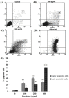
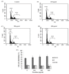
Results: the viable cell number of treated HSC-3 cells was decreased. Moreover, treated cells were arrested in the G0/G1 phase. Annexin V/PI staining revealed that fucoidan could induce apoptosis in HSC-3 cells. Western blot analysis suggested the up-regulation of apoptotic markers including cleaved caspase-3, cleaved PARP, Bax, and autophagic markers including LC3-II and Beclin-1 but down-regulation of anti-apoptotic markers, Bcl-2. Fucoidan could disturb ΔΨm and induce chromatin condensation with nuclear fragmentation.
Conclusion: fucoidan has potential in anticancer properties against HSC-3 cells manifested by the induction of apoptosis, cell cycle arrest, and autophagy.<br />.
Keywords: Apoptosis; Autophagy; Cell cycle; Fucoidan; HSC-3 oral squamous cell carcinoma.
Figures


Similar articles
-
Anticancer Activity of Fucoidan via Apoptosis and Cell Cycle Arrest on Cholangiocarcinoma Cell.Asian Pac J Cancer Prev. 2021 Jan 1;22(1):209-217. doi: 10.31557/APJCP.2021.22.1.209. Asian Pac J Cancer Prev. 2021. PMID: 33507701 Free PMC article.
-
Chloroform Extract of Solanum lyratum Induced G0/G1 Arrest via p21/p16 and Induced Apoptosis via Reactive Oxygen Species, Caspases and Mitochondrial Pathways in Human Oral Cancer Cell Lines.Am J Chin Med. 2015;43(7):1453-69. doi: 10.1142/S0192415X15500822. Epub 2015 Oct 18. Am J Chin Med. 2015. PMID: 26477797
-
Fucoidan induces G1 phase arrest and apoptosis through caspases-dependent pathway and ROS induction in human breast cancer MCF-7 cells.J Huazhong Univ Sci Technolog Med Sci. 2013 Oct;33(5):717-724. doi: 10.1007/s11596-013-1186-8. Epub 2013 Oct 20. J Huazhong Univ Sci Technolog Med Sci. 2013. PMID: 24142726
-
The Therapeutic Potential of the Anticancer Activity of Fucoidan: Current Advances and Hurdles.Mar Drugs. 2021 May 10;19(5):265. doi: 10.3390/md19050265. Mar Drugs. 2021. PMID: 34068561 Free PMC article. Review.
-
Brown algal bioactive molecules: a new frontier in oral cancer treatment.Nat Prod Res. 2025 May;39(10):3005-3007. doi: 10.1080/14786419.2024.2367013. Epub 2024 Jun 17. Nat Prod Res. 2025. PMID: 38884119 Review.
Cited by
-
Type I Cystatin Derived from Fasciola gigantica Suppresses Macrophage-Mediated Inflammatory Responses.Pathogens. 2023 Mar 1;12(3):395. doi: 10.3390/pathogens12030395. Pathogens. 2023. PMID: 36986318 Free PMC article.
-
Brown Algae-Derived Fucoidan Exerts Oxidative Stress-Dependent Antiproliferation on Oral Cancer Cells.Antioxidants (Basel). 2022 Apr 26;11(5):841. doi: 10.3390/antiox11050841. Antioxidants (Basel). 2022. PMID: 35624705 Free PMC article.
-
Ethyl Acetate Extract of Halymenia durvillei Induced Apoptosis, Autophagy, and Cell Cycle Arrest in Colorectal Cancer Cells.Prev Nutr Food Sci. 2023 Mar 31;28(1):69-78. doi: 10.3746/pnf.2023.28.1.69. Prev Nutr Food Sci. 2023. PMID: 37066031 Free PMC article.
-
Anti-Inflammatory Effect of Garcinol Extracted from Garcinia dulcis via Modulating NF-κB Signaling Pathway.Nutrients. 2023 Jan 22;15(3):575. doi: 10.3390/nu15030575. Nutrients. 2023. PMID: 36771283 Free PMC article.
-
Anticancer Activity of Fucoidan via Apoptosis and Cell Cycle Arrest on Cholangiocarcinoma Cell.Asian Pac J Cancer Prev. 2021 Jan 1;22(1):209-217. doi: 10.31557/APJCP.2021.22.1.209. Asian Pac J Cancer Prev. 2021. PMID: 33507701 Free PMC article.
References
-
- Alekseyenko T, Zhanayeva SY, Venediktova A, et al. Antitumor and antimetastatic activity of fucoidan, a sulfated polysaccharide isolated from the Okhotsk Sea Fucus evanescens brown alga. Bull Exp Biol Med. 2007;143:730–2. - PubMed
-
- Asanuma K., Tanida I, Shirato I, et al. MAP-LC3, a promising autophagosomal marker, is processed during the differentiation and recovery of podocytes from PAN nephrosis. FASEB J. 2003;17:1165–7. - PubMed
-
- Atashrazm F, Lowenthal RM, Woods GM, et al. Fucoidan suppresses the growth of human acute promyelocytic leukemia cells in vitro and in vivo. J Cell Physiol. 2016;231:688–97. - PubMed
-
- Banafa AM, Roshan S, Liu YY, et al. Fucoidan induces G1 phase arrest and apoptosis through caspases-dependent pathway and ROS induction in human breast cancer MCF-7 cells. J Huazhong Univ Sci Technol Med Sci. 2013;33:717–24. - PubMed
MeSH terms
Substances
LinkOut - more resources
Full Text Sources
Other Literature Sources
Medical
Research Materials
Miscellaneous

