. 2020 Oct;19(10):e13221.
doi: 10.1111/acel.13221. Epub 2020 Aug 28.

GSK3-ARC/Arg3.1 and GSK3-Wnt signaling axes trigger amyloid-β accumulation and neuroinflammation in middle-aged Shugoshin 1 mice

Affiliations

GSK3-ARC/Arg3.1 and GSK3-Wnt signaling axes trigger amyloid-β accumulation and neuroinflammation in middle-aged Shugoshin 1 mice

Chinthalapally V Rao et al. Aging Cell. 2020 Oct.

Abstract

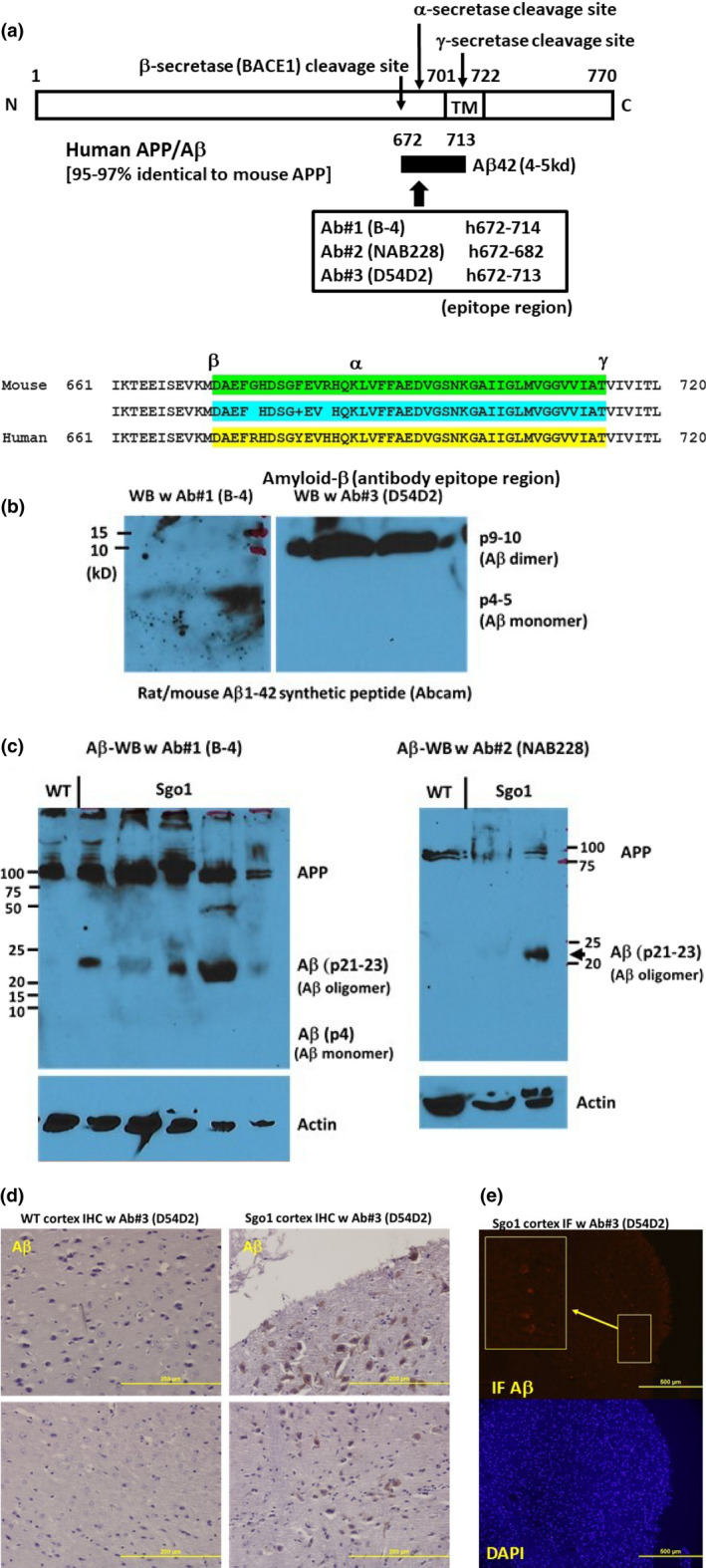
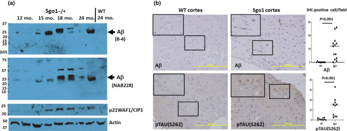
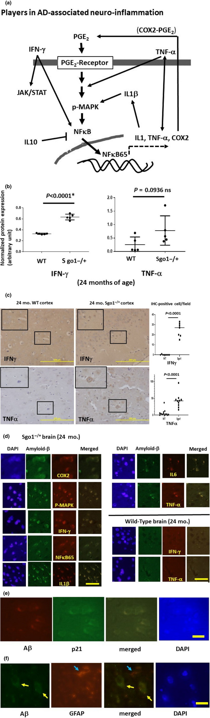
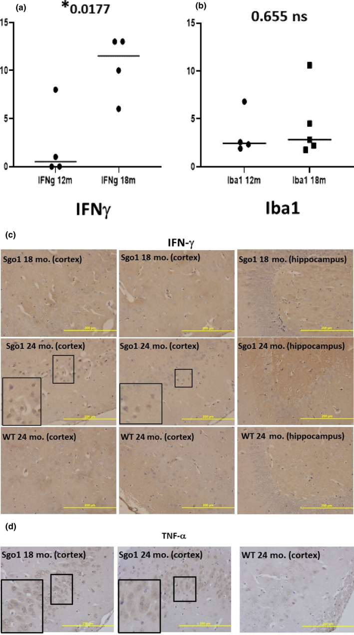
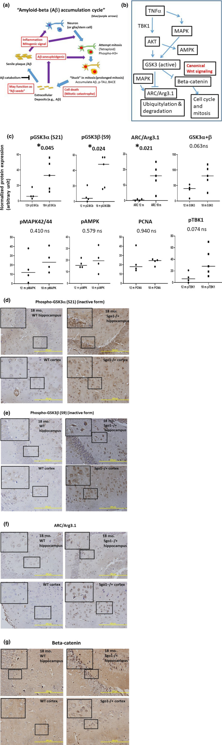
The cerebral amyloid-β accumulation that begins in middle age is considered the critical triggering event in the pathogenesis of late-onset Alzheimer's disease (LOAD). However, the molecular mechanism remains elusive. The Shugoshin 1 (Sgo1-/+ ) mouse model, a model for mitotic cohesinopathy-genomic instability that is observed in human AD at a higher rate, showed spontaneous accumulation of amyloid-β in the brain at old age. With the model, novel insights into the molecular mechanism of LOAD development are anticipated. In this study, the initial appearance of cerebral amyloid-β accumulation was determined as 15-18 months of age (late middle age) in the Sgo1-/+ model. The amyloid-β accumulation was associated with unexpected GSK3α/β inactivation, Wnt signaling activation, and ARC/Arg3.1 accumulation, suggesting involvement of both the GSK3-Arc/Arg3.1 axis and the GSK3-Wnt axis. As observed in human AD brains, neuroinflammation with IFN-γ expression occurred with amyloid-β accumulation and was pronounced in the aged (24-month-old) Sgo1-/+ model mice. AD-relevant protein panels (oxidative stress defense, mitochondrial energy metabolism, and β-oxidation and peroxisome) analysis indicated (a) early increases in Pdk1 and Phb in middle-aged Sgo1-/+ brains, and (b) misregulations in 32 proteins among 130 proteins tested in old age. Thus, initial amyloid-β accumulation in the Sgo1-/+ model is suggested to be triggered by GSK3 inactivation and the resulting Wnt activation and ARC/Arg3.1 accumulation. The model displayed characteristics and affected pathways similar to those of human LOAD including neuroinflammation, demonstrating its potential as a study tool for the LOAD development mechanism and for preclinical AD drug research and development.

Keywords: Shugoshin1; amyloid-β; cohesinopathy; genomic instability; late-onset Alzheimer's disease; mitosis; mouse model; neuroinflammation.


Conflict of interest statement

The authors declare no conflicts of interest.

Figures

FIGURE 1
FIGURE 1
Cerebral amyloid‐β is accumulated in aged (24 month‐old) Sgo1−/+ mice. (a) Human/mouse APP structure. Anti‐amyloid‐β antibodies used for this study were generated against human amyloid‐β, which is 97% identical to mouse amyloid‐β. In humans, B‐4 and NAB228 recognize both APP and amyloid‐β, while D54D2 preferentially recognizes amyloid‐β. (b) Synthetic rat/mouse amyloid‐β1−42 peptide was recognized by anti‐ amyloid‐β antibodies. Amyloid‐β can form SDS‐resistant oligomers that may expose epitope regions differently. B‐4 antibody preferentially recognized monomer (p4‐5), while D54D2 recognized dimer (p9‐10). (c) Twenty‐four‐month‐old Sgo1−/+ brain extracts showed amyloid‐β p21‐23. Both anti‐amyloid‐β antibodies, B‐4 (left panel) and NAB228 (right panel), detected APP and amyloid‐β p21‐23 in immunoblots. Immunoblots of extracts from age‐matched wild‐type mice detected only APP. (d) Twenty‐four ‐month‐old Sgo1−/+ brain showed amyloid‐β accumulation in IHC. Control age‐matched wild‐type mice did not show IHC‐positive staining. (e) Twenty‐four‐month‐old Sgo1−/+ brain showed amyloid‐β accumulation in IF. IF showed positive signals in Sgo1−/+ brain, consistent with IHC results. Enlarged panel shows the signals from cell bodies. Control wild‐type mice did not show clear signals with equalized image acquisition settings (not shown)
FIGURE 2
FIGURE 2
Late‐onset nature of cerebral amyloid‐β accumulation in Sgo1−/+ mice. (a) Cerebral amyloid‐β accumulated by 18 months of age in Sgo1−/+ mice. Extracts were prepared from brains from Sgo1−/+ mice at 12, 15, 18, and 24 months of age. Extracts were probed for amyloid‐β (with B‐4, top panel), amyloid‐β (with NAB228, middle panel), aging marker p21WAF1/CIP1, and actin (loading control). (b) amyloid‐β accumulation in Sgo1−/+ brain cortex occurred in cell bodies. Amyloid‐β IHC with D54D2 antibody for 18‐month‐old Sgo1−/+ brain showed positive signals. However, even at 24 months of age, wild‐type mice showed no positive signals. Bar = 200 µm. Marked fields are enlarged to show IHC details; Aβ and p‐TAU (S262) were observed in cell bodies in Sgo1−/+ cortex, while no signal was seen in wild‐type. IHC‐positive cells were counted in pictures of cortex (N = 10–13), and the number of IHC‐positive cells per field is presented. (c) p‐TAUS262 staining appeared in 18‐month‐old Sgo1−/+ brain. As with amyloid‐β, even at 24 months of age, wild‐type mice showed no positive staining
FIGURE 3
FIGURE 3
Neuroinflammation in Sgo1−/+ mice. (a) Players in AD‐associated neuroinflammation. For this analysis, we prioritized neuroinflammation markers that are known to be upregulated (i.e., IFN‐γ, TNF‐α, NFκB65, IL1‐β, IL6, and COX‐2) or downregulated (i.e., IL10) in the brains of human patients with AD. Overall, the neuroinflammation markers are proposed to form feedback loops. One messenger is Prostaglandin E2 (PGE2), which binds to the receptor and activates a cascade, leading to MAPK phosphorylation and activation of the NFκB 65kd subunit. The NFκB 65kd subunit translocates to the nucleus and activates downstream transcriptions of other inflammatory mediators, including IL1, TNF‐α, and COX‐2. COX‐2, in turn, generates PGE2. Inflammatory cytokine IL1‐β activates COX‐2. TNF‐α is also thought to activate p38MAPK. Inflammatory cytokine Interferon‐γ (IFN‐γ) leads to activations of JAK/STAT and NFκB. However, interpreting the role of neuroinflammation markers may not be straightforward. (b) AD‐associated neuroinflammation marker IFN‐γ was upregulated in aged, amyloid‐β‐accumulating Sgo1−/+ brains (p < 0.0001). Another marker, TNF‐α, was consistently upregulated in Sgo1−/+, showing the dual presence of IFN‐γ and TNF‐α in Sgo1−/+ brain. However, inconsistent expression of TNF‐α in wild‐type control mice led to a non‐significant p‐value (p = .0936). (c) IFN‐γ and TNF‐α in Sgo1−/+ brain. As suggested by immunoblot results in (b), IFN‐γ and TNF‐α were both detected in aged Sgo1−/+ brain. Also consistent with immunoblots, IHC of wild‐type controls showed much less IFN‐γ and TNF‐α. IFN‐γ and TNF‐α were observed in cell bodies in Sgo1−/+ cortex, while no signal was seen in wild‐type (enlarged panels). IHC‐positive cells were counted in pictures of cortex (N = 7–12), and the number of IHC‐positive cells per field is presented. (d) Accumulating amyloid‐β and neuroinflammation markers (COX2, p‐MAPK, IFN‐γ, NFκB65, IL1‐β, IL6, and TNF‐α) co‐localized in Sgo1−/+ brain cortex cells. Age‐matched wild‐type mice showed little amyloid‐β, and co‐localization was not observed. Scale bar (yellow):50 µm. (e) Amyloid‐β and p21 co‐localized in Sgo1−/+ brain cortex cells. Scale bar (yellow):20 µm. (f) Amyloid‐β and Glial fibrillary acidic protein (GFAP) did not co‐localize in Sgo1−/+ brain cortex cells. GFAP is a marker for astrocytes and ependymal cells. Aβ‐positive cells (yellow arrow) and GFAP‐positive cells (blue arrow) are different cells. Scale bar (yellow): 20 µm
FIGURE 4
FIGURE 4
IFN‐γ ‐mediated neuroinflammation was concurrent with, but did not precede, Aβ accumulation. (a) IFN‐γ increased by age 18 months in Sgo1−/+. The amount of IFN‐γ in Sgo1−/+ brain was low at 12 months of age, but increased by 18 months of age. Protein amounts, measured by immunoblot and normalized with actin amount, are plotted and compared. (b) Microglia infiltration may not accompany IFN‐γ increase at the 12‐18 month transition. Microglia marker Iba1 (measured as in (a)) did not show a significant increase in 18‐month‐old Sgo1−/+, suggesting that infiltration of microglia may not be significantly increased at this age. (c) IFN‐γ localized in nucleo‐cytoplasm in aged Sgo1−/+. Distinct localization of IFN‐γ in nucleo‐cytoplasm was observed only in aged (24‐month) samples and was not observed in 18‐month samples. Marked fields are enlarged to show IHC details. IFN‐γ was observed in cell bodies in 24‐month Sgo1−/+ cortex, while no localized signal was seen in 18‐month Sgo1−/+ brain samples. Wild‐type controls did not show IFN‐γ at any age. (d) TNF‐α localization in cytoplasm and in nucleo‐cytoplasm. Distinct cell body localization of TNF‐α was observed in Sgo1−/+ cortex at both 18 and 24 months of age. At 18 months, cytoplasmic staining was evident (enlarged panel), while at 24 months, diffused staining in both nucleoplasm and cytoplasm was common
FIGURE 5
FIGURE 5
Inhibition of GSK3 α and β, accumulation of ARC/Arg3.1, and activation of Wnt signaling in amyloid‐accumulating Sgo1−/+. (a) The “amyloid‐β accumulation cycle” hypothesis (Rao et al., 2020). The “amyloid‐β accumulation cycle” hypothesis purports the occurrence of vicious cycles of events leading to amyloid‐β accumulation (see Introduction). Among a few mysteries in the hypothesis is the growth signaling driving the amyloid‐β accumulation cycle. (b) Key growth signaling pathways that are misregulated in human AD patients. AKT, AMPK, MAPK, and GSK3 are among the growth signaling misregulated in human AD and proposed to be involved in the disease process. GSK3 targets ARC/Arg3.1 and β‐catenin with ubiquitylation‐mediated proteolysis. (c) Phosphorylated GSK3 α and β (inactive forms) increased in Aβ‐accumulating Sgo1−/+. We tested components of the growth signaling in (b). Amounts of pGSK3α (S21) and pGSK3β (S9), inactive forms of GSK3, increased significantly in Aβ‐accumulating Sgo1−/+, while the total amount of GSK3 showed only a minor change. Consistently, ARC/Arg3.1 amount also significantly increased. pMAPK42/44, pAMPK, PCNA, and pTBK1 did not show significant change. (d) Nuclear accumulation of pGSK3α (S21) in Sgo1−/+. Consistent with immunoblots in (c) Aβ‐accumulating Sgo1−/+ showed accumulation of pGSK3α in the nucleus, both in the hippocampus and in the cortex. Age‐matched wild‐type showed no such pGSK3α accumulation. Enlarged panels for localization details. (e) Cytoplasmic accumulation of pGSK3β (S9) in Sgo1−/+. Aβ ‐accumulating Sgo1−/+ showed accumulation of pGSK3β in the cytoplasm, both in the hippocampus and in the cortex. Enlarged panels for localization details. (f) ARC/Arg3.1 was accumulated in the nucleo‐cytoplasm. ARC/Arg3.1 was accumulated in the nucleo‐cytoplasm, in both the hippocampus and the cortex, in Sgo1−/+. Enlarged panels for localization details. In wild‐type, IHC signals for ARC/Arg3.1 were much weaker, if any. (g) Another GSK3 target β‐catenin was enriched in the nuclei of Sgo1−/+, indicating Wnt signaling activation. Consistent with GSK3 inactivation, nuclear translocation of β‐catenin, a sign of canonical Wnt signaling activation and cell fate toward cell cycle and mitosis, was observed in Sgo1−/+ as distinct nucleo‐cytoplasmic signals in both the hippocampus and the cortex. β‐catenin IHC signals in wild‐type were weak, if any (enlarged panels)
FIGURE 6
FIGURE 6
Protein misregulations in Sgo1−/+ in human AD‐relevant proteins (i.e., antioxidant proteins, mitochondrial energy metabolism proteins, and β‐oxidation and peroxisome proteins). (a) Examples of proteins indicating an Sgo1−/+‐specific increase in 24‐month‐old brains. Hspd1 (Heat Shock Protein Family D [Hsp60] Member 1) is a mitochondrial chaperone. Gpi (Glucose‐6‐phosphate isomerase) and Fh1 (Fumarate Hydratase) are involved in energy generation. (b) Examples of proteins indicating both an Sgo1−/+‐specific increase at 24 months and age‐dependent increase in Sgo1−/+ (12 months vs. 24 months). Idh1 (Isocitrate Dehydrogenase [NADP(+)] 1, Cytosolic), and Idh2 (Isocitrate Dehydrogenase [NADP(+)] 2, Mitochondrial) are involved in energy generation. Aco2 (aconitase 2, mitochondrial) localizes in mitochondria and is a part of the TCA cycle. (c) Prohibitin (Phb) and Phosphoinositide‐dependent protein kinase 1(Pdk1) uniquely indicated increases in middle‐aged (12‐month‐old) Sgo1−/+ mice, preceding amyloid‐β accumulation at 24 months. Phb and Pdk1 are also progressively decreased in human olfactory bulb‐AD proteomic analysis (see text). (d) The majority of misregulations were increases at old age, while Phb2 (Prohibitin2), Fabp3 (Fatty Acid‐binding Protein 3), and nnt (Nicotinamide Nucleotide Transhydrogenase) showed decreases. Many of these proteins are also misregulated in human AD (see text)

Similar articles

Cited by

References

    1. Andrew, R. J. , Kellett, K. A. , Thinakaran, G. , & Hooper, N. M. (2016). A Greek tragedy: The growing complexity of Alzheimer amyloid precursor protein proteolysis. Journal of Biological Chemistry, 291(37), 19235–19244. - PMC - PubMed
    1. Andriani, G. A. , Vijg, J. , & Montagna, C. (2017). Mechanisms and consequences of aneuploidy and chromosome instability in the aging brain. Mechanisms of Ageing and Development, 161(Pt A), 19–36. - PMC - PubMed
    1. Avrahami, L. , Farfara, D. , Shaham‐Kol, M. , Vassar, R. , Frenkel, D. , & Eldar‐Finkelman, H. (2013). Inhibition of glycogen synthase kinase‐3 ameliorates β‐amyloid pathology and restores lysosomal acidification and mammalian target of rapamycin activity in the Alzheimer disease mouse model: in vivo and in vitro studies. Journal of Biological Chemistry, 288(2), 1295–1306. 10.1074/jbc.M112.409250 - DOI - PMC - PubMed
    1. Bajic, V. , Spremo‐Potparevic, B. , Zivkovic, L. , Isenovic, E. R. , & Arendt, T. (2015). Cohesion and the aneuploid phenotype in Alzheimer's disease: A tale of genome instability. Neuroscience and Biobehavioral Reviews, 55, 365–374. - PubMed
    1. Bakhoum, S. F. , Ngo, B. , Laughney, A. M. , Cavallo, J. A. , Murphy, C. J. , Ly, P. , … Cantely, L. C. (2018). Chromosomal instability drives metastasis through a cytosolic DNA response. Nature, 553(7689), 467–472. 10.1038/nature25432 - DOI - PMC - PubMed

Publication types