. 2020 Aug 28;26(6):613-623.
doi: 10.3171/2020.5.PEDS20124. Print 2020 Dec 1.

Acute brain inflammation, white matter oxidative stress, and myelin deficiency in a model of neonatal intraventricular hemorrhage

Affiliations

Acute brain inflammation, white matter oxidative stress, and myelin deficiency in a model of neonatal intraventricular hemorrhage

Danielle S Goulding et al. J Neurosurg Pediatr. .

Abstract

Objective: Neonatal intraventricular hemorrhage (IVH) leads to posthemorrhagic hydrocephalus (PHH), brain injury, and long-term disability. Current therapy for IVH is based on treating PHH but does not address the underlying brain injury. In order to develop pharmacological treatment for IVH, there must be a better understanding of the underlying pathology of this disease. This study was designed to determine the time course of the acute inflammation and oxidative stress that may underlie the progressive pathology of IVH. The authors sought to understand the temporal relationships among inflammation, oxidative stress, and white matter pathology in a rat model of IVH.

Methods: A rat model of IVH consisting of hemoglobin injection into the lateral ventricle was used. Tissue was analyzed via biochemical and histological methods to map the spatiotemporal distribution of innate immune activation and oxidative stress. White matter was quantified using both immunohistochemistry and Western blot for myelin basic protein (MBP) in the corpus callosum.

Results: IVH led to acute induction of inflammatory cytokines, followed by oxidative stress. Oxidative stress was concentrated in white matter, adjacent to the lateral ventricles. Animals with IVH initially gained weight at a lower rate than control animals and had larger ventricles and less MBP than control animals.

Conclusions: Experimental IVH induces global inflammation throughout the brain and oxidative stress concentrated in the white matter. Both of these phenomena occur early after IVH. This has implications for human neonates with immature white matter that is exquisitely sensitive to inflammation and oxidative stress. Antiinflammatory or antioxidant therapy for IVH may need to be initiated early in order to protect developing white matter.

Keywords: CCR2; hydrocephalus; intraventricular hemorrhage; microglia.


Conflict of interest statement

The authors report no conflict of interest concerning the materials or methods used in this study or the findings specified in this paper.

Figures

FIG. 1.
FIG. 1.
Weight gain after IVH induction. Twenty-four hours after IVH induction, control animals had gained 17.8% ± 6.3% of their preinjection body weight and IVH animals had gained 8.0% ± 5.6% (p = 0.004, n = 47–59 per group). Forty-eight hours after IVH induction, control animals had gained 34.0% ± 8.1% of their preinjection body weight and IVH animals had gained 23.6% ± 8.8% (p = 0.03, n = 20–25 per group). Seven days (7D) after injection, there was no significant difference in weight gained from the day of injection in control and IVH animals (n = 12–16 per group). *p < 0.05.
FIG. 2.
FIG. 2.
Cytokine measurements in the hemisphere ipsilateral to hemoglobin or saline injection. A: TNFα was significantly increased at 3 hours after IVH induction (5.0 ± 2.1 vs 0.02 ± 0.003 pg/ml/mg in controls, p < 0.0001, n = 5–6 per group). There was a trend toward increased TNFα 6 hours after IVH (1.1 ± 0.4 vs 0.03 ± 0.003 pg/ml/mg in controls, not significant, n = 6 per group). B: CXCL1 was increased at 3 and 6 hours after IVH (14.4 ± 2.6 vs 0.2 ± 0.08 pg/ml/mg in controls at 3 hours, p < 0.0001, n = 5–6 per group; 14.1 ± 3.1 vs 0.2 ± 0.04 pg/ml/mg in controls at 6 hours, p < 0.0001, n = 6 per group). C: IL-1β was increased at 3 and 6 hours after IVH (1.03 ± 0.14 vs 0.73 ± 0.8 pg/ml/mg in controls at 3 hours, p = 0.0006, n = 5–6 per group; 1.04 ± 0.14 vs 0.78 ± 0.01 pg/ml/mg in controls at 6 hours, p = 0.001, n = 6 per group). D: IL-6 was increased at 3 and 6 hours after IVH (7.1 ± 1.3 vs 3.5 ± 0.3 pg/ml/mg in controls at 3 hours, p = 0.0014, n = 5–6 per group; 14.1 ± 3.2 vs 3.6 ± 0.2 pg/ml/mg in controls at 6 hours, p < 0.0001, n = 6 per group). *p < 0.05.
FIG. 3.
FIG. 3.
Oxidative stress after IVH in the hemisphere ipsilateral to saline or hemoglobin injection. There was a significant increase in 4HNE 24 hours after IVH induction (0.76 ± 0.3 normalized signal vs 0.2 ± 0.06 normalized signal in controls at 3 hours, p < 0.0001, n = 5–6 per group except the 7D control group in which n = 1). *p < 0.05.
FIG. 4.
FIG. 4.
Microglia and macrophages increase after IVH. Quantitative analysis of IBA1 immunohistochemistry in the cerebral cortex (A) showed no effect of IVH on IBA1 labeling (B). Quantitative analysis of IBA1 labeling in white matter (C) showed a significant effect of IVH at 3 and 24 hours after IVH (91.3 ± 45.2 vs 17.5 ± 8.4 cells/mm2 in controls at 3 hours, p = 0.002, n = 3–5 per group; 172.3 ± 65.4 vs 75.2 ± 30.6 cells/mm2 in controls at 24 hours, p = 0.03, n = 3–5 per group; D). Data from hemisphere ipsilateral to saline or hemoglobin injection. *p < 0.05.
FIG. 5.
FIG. 5.
Macrophage reactivity increases after IVH. Quantitative analysis of CD68 immunohistochemistry in the cerebral cortex (A) showed a significant effect of IVH 3 hours, 24 hours, and 7 days (7D) after induction (88.5 ± 35.6 vs 10.9 ± 9.2 cells/mm2 in controls at 3 hours, p = 0.004, n = 3–5 per group; 19.7 ± 8.5 vs 6.4 ± 1.2 cells/mm2 in controls at 24 hours, p = 0.01, n = 3–5 per group; 0.8 ± 0.5 vs 0.2 ± 0.1 cells/mm2 in controls at 7D, p = 0.02, n = 5–6 per group; B). Quantitative analysis of CD68 labeling in white matter (C) showed a significant effect of IVH 3 and 24 hours after induction (187.5 ± 60.3 vs 42.0 ± 19.7 cells/mm2 in controls at 3 hours, p = 0.002, n = 3–5 per group; 205.8 ± 60.4 vs 101.6 ± 23.4 cells/mm2 in controls at 24 hours, p = 0.02, n = 3–5 per group; D). Data from hemisphere ipsilateral to saline or hemoglobin injection. *p < 0.05.
FIG. 6.
FIG. 6.
CCL2/CCR2 is induced in myeloid cells after IVH. A: rtPCR of the myeloid fraction of whole brain revealed a significant increase in CCL2 induction 3 hours after IVH (4.2 log2 fold change from saline controls at 3 hours, p < 0.001, n = 4 per group). There was a significant decrease in CCL2 induction 72 hours after IVH (−1.9 log2 fold change from controls at 3 hours, p = 0.01, n = 4 per group). B: CCR2 was increased at 3, 24, and 72 hours after IVH (2.2 log2 fold change, 3.4 log2 fold change, and 1.8 log2 fold change from saline control at 3 hours, p < 0.0001, p < 0.0001, and p = 0.0006, respectively). **p < 0.05.
FIG. 7.
FIG. 7.
IVH selectively increases oxidative stress in the white matter of brain ipsilateral to injury. Quantitative immunohistochemistry for 15A3 in the cerebral cortex (A) revealed no effect of IVH (B). Quantitative immunohistochemistry for 15A3 in white matter (C) revealed an increase in oxidative stress 24 hours (31.3% ± 7.8% positive area vs 7.7% ± 4.3% positive area in controls, p = 0.01, n = 3–4 per group) and 7 days (7D) after IVH (10.2% ± 4.5% positive area vs 5.5% ± 0.4% positive area in controls, p = 0.002, n = 5–6 per group; D). *p < 0.05.
FIG. 8.
FIG. 8.
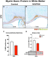
IVH leads to a reduction in white matter. Immunohistochemistry for MBP (A) at 37 days (37D) postinjection showed a trend toward less MBP labeling in animals that had IVH (B; 49.0% ± 21.4% positive area after IVH vs 68.6% ± 10.9% positive area in controls, p = 0.12, n = 4–9 per group; C). Western blot of MBP performed on isolated corpus callosum revealed a significant reduction in MBP 37D after IVH (D; 0.46 ± 0.07 normalized MBP signal after IVH vs 0.63 ± 0.11 normalized signal in controls, p = 0.01, n = 5–6 per group). All Western blot data are shown. *p < 0.05.
FIG. 9.
FIG. 9.
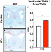
Ventriculomegaly develops after IVH. The ratio of ventricular width/total brain width (A) was increased in animals with IVH 37D after IVH induction (B; 0.4 ± 0.06 vs 0.3 ± 0.02 for controls, p = 0.003, n = 7–9 per group; C). *p < 0.05.

References

    1. Christian EA, Jin DL, Attenello F, et al. Trends in hospitalization of preterm infants with intraventricular hemorrhage and hydrocephalus in the United States, 2000–2010. J Neurosurg Pediatr. 2016;17(3):260–269. - PubMed
    1. Martin JA, Kung HC, Mathews TJ, et al. Annual summary of vital statistics: 2006. Pediatrics. 2008;121(4):788–801. - PubMed
    1. Volpe JJ. Brain injury in premature infants: a complex amalgam of destructive and developmental disturbances. Lancet Neurol. 2009;8(1):110–124. - PMC - PubMed
    1. Mukerji A, Shah V, Shah PS. Periventricular/intraventricular hemorrhage and neurodevelopmental outcomes: a meta-analysis. Pediatrics. 2015;136(6):1132–1143. - PubMed
    1. Kahle KT, Kulkarni AV, Limbrick DD, Jr, Warf BC. Hydrocephalus in children. Lancet. 2016;387(10020):788–799. - PubMed