. 2020 Aug 26;20(17):4805.
doi: 10.3390/s20174805.

Wearable Tendon Kinetics

Affiliations

Wearable Tendon Kinetics

Sara E Harper et al. Sensors (Basel). .

Abstract

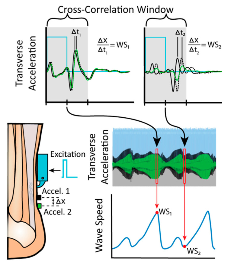
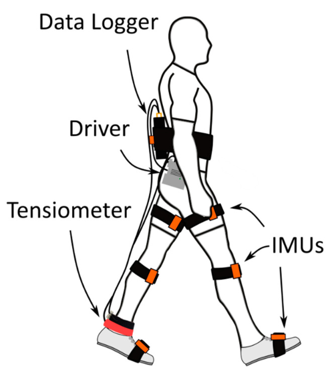
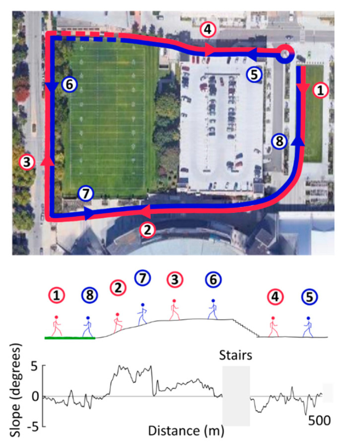
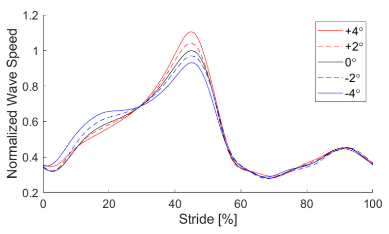
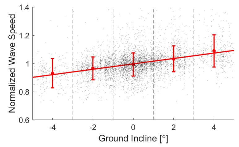
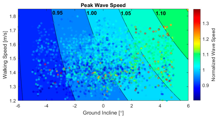
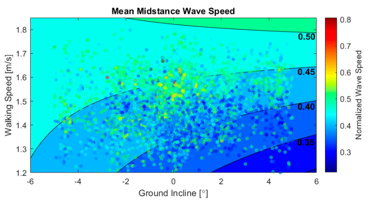
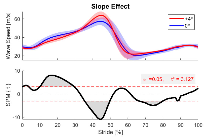
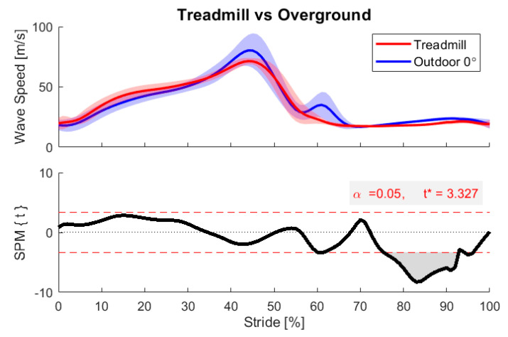
This study introduces a noninvasive wearable system for investigating tendon loading patterns during outdoor locomotion on variable terrain. The system leverages shear wave tensiometry, which is a new approach for assessing tendon load by tracking wave speed within the tissue. Our wearable tensiometry system uses a battery-operated piezoelectric actuator to induce micron-scale shear waves in a tendon. A data logger monitors wave propagation by recording from two miniature accelerometers mounted on the skin above the tendon. Wave speed is determined from the wave travel time between accelerometers. The wearable system was used to record Achilles tendon wave speed at 100 Hz during 1-km outdoor walking trials in nine young adults. Inertial measurement units (IMUs) simultaneously monitored participant position, walking speed, and ground incline. An analysis of 5108 walking strides revealed the coupled biomechanical effects of terrain slope and walking speed on tendon loading. Uphill slopes increased the tendon wave speed during push-off, whereas downhill slopes increased tendon wave speeds during early stance braking. Walking speed significantly modulated peak tendon wave speed on uphill slopes but had less influence on downhill slopes. Walking speed consistently induced greater early stance wave speeds for all slopes. These observations demonstrate that wearable shear wave tensiometry holds promise for evaluating tendon tissue kinetics in natural environments and uncontrolled movements. There are numerous practical applications of wearable tensiometry spanning orthopedics, athletics, rehabilitation, and ergonomics.

Keywords: Achilles; field-based measurement; locomotion; muscle-tendon mechanics; noninvasive; shear wave tensiometry.


Conflict of interest statement

D.T. is a co-inventor of a patent on the shear wave tensiometry technology (US10631775). The funders had no role in the design of the study; in the collection, analyses, or interpretation of data; in the writing of the manuscript, or in the decision to publish the results.

Figures

Figure 1
Figure 1
The tapper mechanism was driven by a 100 Hz pulse wave (2 ms pulse, the time required for one wave to dissipate) excitation signal. The impulsive tap induces a shear wave in the Achilles tendon that is recorded as it passes two distal accelerometers mounted 8 mm (Δx) apart. Cross correlation of the accelerometer signals in a 4-ms window after the excitation onset was used to compute the wave travel time (Δt), which in turn was used to compute the wave speed (WS).
Figure 2
Figure 2
Portable instrumentation used in the outdoor walking tests. A shear wave tensiometer was mounted over the right Achilles tendon, wired to a battery-powered piezo driver and data logger mounted on the waist and low back. Stride length, walking speed, and position along the prescribed walking course were recorded from the XSens lower-body IMU model (XSens MVN Analyze software; IMUs shown in orange).
Figure 3
Figure 3
Aerial view of the 1-km outdoor walking course, including ground inclines, declines, and stairs. Four of the participants started near the edge of the grass and walked a lap according to the figure shown. They then turned around and walked the same course in the opposite direction. The remaining five participants walked the course in the opposite direction (5 to 8, then 1 to 4). The ground incline data when traversing the course in clockwise direction is shown in the bottom trace. Stairs, shown in broken lines (top) or hidden (bottom), were excluded from the analysis due to faulty data and behavioral variability.
Figure 4
Figure 4
Ensembled Achilles tendon wave speed patterns at binned inclines. Normalization is to mean peak wave speed in the level walking condition for each participant. Peak wave speed of each stride was aligned to 45% of the stride to approximate a traditional gait cycle for visualization [49].
Figure 5
Figure 5
A regression of peak loading against ground incline yielded a positive association (p < 10−6). Means and standard deviations for each binned incline demonstrate the trend, while the spread of data shows the remaining variability in the data. Regression results for individual subjects are shown in Appendix B.
Figure 6
Figure 6
Scatter plot of peak wave speed vs. ground incline and walking speed for all 5108 strides. Data are plotted on top of contours of constant peak wave speed from a multiple linear regression with interaction.
Figure 7
Figure 7
Scatter plot of the mean midstance wave speed vs. ground incline and walking speed for all 5108 strides. Data are plotted on top of contours of the constant midstance wave speed from a multiple linear regression with interaction.
Figure 8
Figure 8
Statistical parametric mapping (SPM) between level walking (0° incline bin) and steep inclined walking (+4° bin) for one example participant. SPM revealed regions of significant difference in the two conditions (gray regions). For incline walking, meaningful differences were detected in early stance (around 20% stride) and at the push-off peak (45% stride).
Figure 9
Figure 9
Statistical parametric mapping between in-lab and outdoor walking (0° incline bin) for one participant. SPM revealed a region of significant difference between these conditions (gray region), here in the swing phase. Greater variability was observed in the outdoor condition.

References

    1. Chambers R., Gabbett T.J., Cole M.H., Beard A. The Use of Wearable Microsensors to Quantify Sport-Specific Movements. Sports Med. 2015;45:1065–1081. doi: 10.1007/s40279-015-0332-9. - DOI - PubMed
    1. Li R.T., Kling S.R., Salata M.J., Cupp S.A., Sheehan J., Voos J.E. Wearable Performance Devices in Sports Medicine. Sports Health. 2016;8:74–78. doi: 10.1177/1941738115616917. - DOI - PMC - PubMed
    1. Peppoloni L., Filippeschi A., Ruffaldi E., Avizzano C.A. (WMSDs Issue) A Novel Wearable System for the Online Assessment of Risk for Biomechanical Load in Repetitive Efforts. Int. J. Ind. Ergon. 2014;52:1. doi: 10.1016/j.ergon.2015.07.002. - DOI
    1. Alberto R., Draicchio F., Varrecchia T., Silvetti A., Iavicoli S. Wearable Monitoring Devices for Biomechanical Risk Assessment at Work: Current Status and Future Challenges—A Systematic Review. Int. J. Environ. Res. Public Health. 2018;15:2001. doi: 10.3390/ijerph15092001. - DOI - PMC - PubMed
    1. Conforti I., Mileti I., Del Prete Z., Palermo E. Assessing Ergonomics and Biomechanical Risk in Manual Handling of Loads through a Wearable System; Proceedings of the 2019 IEEE International Workshop on Metrology for Industry 4.0 and IoT, MetroInd 4.0 and IoT; Naples, Italy. 4–6 June 2019; - DOI

LinkOut - more resources