Review
. 2020 Aug 26;9(9):1961.
doi: 10.3390/cells9091961.

Non-Muscle Myosin II in Axonal Cell Biology: From the Growth Cone to the Axon Initial Segment

Affiliations
Review

Non-Muscle Myosin II in Axonal Cell Biology: From the Growth Cone to the Axon Initial Segment

Ana Rita Costa et al. Cells. .

Abstract

By binding to actin filaments, non-muscle myosin II (NMII) generates actomyosin networks that hold unique contractile properties. Their dynamic nature is essential for neuronal biology including the establishment of polarity, growth cone formation and motility, axon growth during development (and axon regeneration in the adult), radial and longitudinal axonal tension, and synapse formation and function. In this review, we discuss the current knowledge on the spatial distribution and function of the actomyosin cytoskeleton in different axonal compartments. We highlight some of the apparent contradictions and open questions in the field, including the role of NMII in the regulation of axon growth and regeneration, the possibility that NMII structural arrangement along the axon shaft may control both radial and longitudinal contractility, and the mechanism and functional purpose underlying NMII enrichment in the axon initial segment. With the advances in live cell imaging and super resolution microscopy, it is expected that in the near future the spatial distribution of NMII in the axon, and the mechanisms by which it participates in axonal biology will be further untangled.

Keywords: actin ring; actomyosin cytoskeleton; axon growth; axon initial segment; axon regeneration; growth cone; non-muscle myosin II.


Conflict of interest statement

The authors declare no conflict of interest.

Figures

Figure 1
Figure 1
Non-muscle myosin II (NMII) structure and regulation. (A) NMII contains two heavy chains (HC), two regulatory light chains (RLCs) (orange) and two essential light chains (ELCs) (yellow). The heavy chain includes the head domain (green), with an actin binding site and an ATPase motor domain, the neck domain to which the ELC and RLC are bound, and the tail domain, with a helical coiled coil rod filament and a non-helical tail. In the absence of RLC phosphorylation, NMII is in an inactive conformation (left). Upon RLC phosphorylation by MLCK or ROCK, for instance, NMII unfolds to generate an active conformation (middle). MLCK phosphorylates RLC on Ser19 and Thr18, depicted as two red circles. The RLC phosphatase MLCP can revert this activation. NMII is then able to assemble into bipolar filaments, which bind to actin (blue) (right). (B) Upon NMII activation, polymerization-competent NMII molecules can form bipolar filaments through electrostatic interactions of their rod domains. Addition of more NMII molecules drives the growth of a bipolar NMII filament. For a matter of simplicity, a smaller number of myosins than that generally found on each side of the filament is drawn. (C) NMII filaments can also form super structures, the NMII stacks. Two models are considered to underlie NMII stack formation: Filament concatenation (left) and filament expansion (right). In the model of filament concatenation, NMII stacks can be formed through concatenation of multiple NMII filaments. In the model of NMII filament expansion, after the formation of the bipolar NMII filament, certain subset of daughter myosins separate themselves from one side of the filament, being each one used as a template. Growth of new templates is driven by addition of NMII filaments.
Figure 2
Figure 2
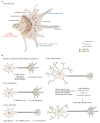
NMII in axon elongation, retraction and regeneration. (A) Schematic representation of the growth cone. The growth cone includes a central domain, mainly consisting of bundled microtubules (grey lines), a transition zone, enriched in actin arcs (blue hemicircular lines) and a peripheral domain, enriched in actin (blue lines) in the form of lamellipodia and filopodia. Actin bundles assemble near the growth cone leading edge, translocate rearward by retrograde flow towards the transition zone (blue dashed arrows in the peripheral domain) and recycle through bundle severing. NMIIA (green) is mainly located within the central domain and NMIIB (red) in the transition zone and peripheral domain. (B) General effect of NMII in neurite extension, axon elongation, retraction and regeneration. During early neurite extension pharmacological inhibition of NMII either directly using blebbistatin, or indirectly through the inhibition of the upstream regulators MLCK or ROCK, promotes axon elongation (upper left panel). When analyzing the effect of different NMII isoforms in axon elongation, in general terms, NMIIB is required for axon elongation (middle left panel) while NMIIA participates in neurite retraction (bottom left panel). In the course of axon regeneration (right panel) NMII inactivation through blebbistatin and/or NMIIA/B downregulation/knockdown, enhances axon regeneration.
Figure 3
Figure 3
Schematic representation of the role of the actomyosin cytoskeleton in the control of axonal contractility. Phosphorylated myosin light chain is highly enriched at the AIS, where it may participate in the regulation of axon diameter during action potential firing. In the axon shaft, circumferential and longitudinal axon tension are also regulated by the actomyosin network. While pMLC forms circular periodic structures colocalizing with membrane periodic skeleton (MPS) actin rings, NMII heavy chains appear mostly distributed as multiple filaments with approximately 300 nm of length along the longitudinal axonal axis, crosslinking adjacent actin rings (probably providing for tension); NMII filaments spanning individual rings may also exist (and provide for contraction).

References

    1. Odronitz F., Kollmar M. Drawing the tree of eukaryotic life based on the analysis of 2,269 manually annotated myosins from 328 species. Genome Biol. 2007;8:R196. doi: 10.1186/gb-2007-8-9-r196. - DOI - PMC - PubMed
    1. Sellers J.R. Myosins: A diverse superfamily. Biochim. Biophys. Acta. 2000;1496:3–22. doi: 10.1016/S0167-4889(00)00005-7. - DOI - PubMed
    1. Pollard T.D., Weihing R.R. Actin and myosin and cell movement. CRC Crit. Rev. Biochem. 1974;2:1–65. doi: 10.3109/10409237409105443. - DOI - PubMed
    1. Pecci A., Ma X., Savoia A., Adelstein R.S. MYH9: Structure, functions and role of non-muscle myosin IIA in human disease. Gene. 2018;664:152–167. doi: 10.1016/j.gene.2018.04.048. - DOI - PMC - PubMed
    1. Vicente-Manzanares M., Ma X., Adelstein R.S., Horwitz A.R. Non-muscle myosin II takes centre stage in cell adhesion and migration. Nat. Rev. Mol. Cell Biol. 2009;10:778–790. doi: 10.1038/nrm2786. - DOI - PMC - PubMed

Publication types

LinkOut - more resources