The Maintenance of Mitochondrial DNA Integrity and Dynamics by Mitochondrial Membranes
- PMID: 32858900
- PMCID: PMC7555930
- DOI: 10.3390/life10090164
The Maintenance of Mitochondrial DNA Integrity and Dynamics by Mitochondrial Membranes
Abstract
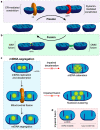
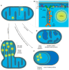
Mitochondria are complex organelles that harbour their own genome. Mitochondrial DNA (mtDNA) exists in the form of a circular double-stranded DNA molecule that must be replicated, segregated and distributed around the mitochondrial network. Human cells typically possess between a few hundred and several thousand copies of the mitochondrial genome, located within the mitochondrial matrix in close association with the cristae ultrastructure. The organisation of mtDNA around the mitochondrial network requires mitochondria to be dynamic and undergo both fission and fusion events in coordination with the modulation of cristae architecture. The dysregulation of these processes has profound effects upon mtDNA replication, manifesting as a loss of mtDNA integrity and copy number, and upon the subsequent distribution of mtDNA around the mitochondrial network. Mutations within genes involved in mitochondrial dynamics or cristae modulation cause a wide range of neurological disorders frequently associated with defects in mtDNA maintenance. This review aims to provide an understanding of the biological mechanisms that link mitochondrial dynamics and mtDNA integrity, as well as examine the interplay that occurs between mtDNA, mitochondrial dynamics and cristae structure.
Keywords: cristae; mitochondria; mitochondrial diseas; mitochondrial fission; mitochondrial fusion; mtDNA.
Conflict of interest statement
The authors declare no conflict of interest. The funders had no role in the design of the study; in the collection, analyses, or interpretation of data; in the writing of the manuscript, or in the decision to publish the results.
Figures


Similar articles
-
Functional Interplay between Cristae Biogenesis, Mitochondrial Dynamics and Mitochondrial DNA Integrity.Int J Mol Sci. 2019 Sep 3;20(17):4311. doi: 10.3390/ijms20174311. Int J Mol Sci. 2019. PMID: 31484398 Free PMC article. Review.
-
Autophagy determines mtDNA copy number dynamics during starvation.Autophagy. 2019 Jan;15(1):178-179. doi: 10.1080/15548627.2018.1532263. Epub 2018 Oct 13. Autophagy. 2019. PMID: 30301401 Free PMC article.
-
Mathematical modeling of the role of mitochondrial fusion and fission in mitochondrial DNA maintenance.PLoS One. 2013 Oct 11;8(10):e76230. doi: 10.1371/journal.pone.0076230. eCollection 2013. PLoS One. 2013. PMID: 24146842 Free PMC article.
-
Mitochondrial Membranes and Mitochondrial Genome: Interactions and Clinical Syndromes.Membranes (Basel). 2022 Jun 15;12(6):625. doi: 10.3390/membranes12060625. Membranes (Basel). 2022. PMID: 35736332 Free PMC article. Review.
-
Integrity of the yeast mitochondrial genome, but not its distribution and inheritance, relies on mitochondrial fission and fusion.Proc Natl Acad Sci U S A. 2015 Mar 3;112(9):E947-56. doi: 10.1073/pnas.1501737112. Epub 2015 Feb 17. Proc Natl Acad Sci U S A. 2015. PMID: 25730886 Free PMC article.
Cited by
-
Growth hormone remodels the 3D-structure of the mitochondria of inflammatory macrophages and promotes metabolic reprogramming.Front Immunol. 2023 Jul 5;14:1200259. doi: 10.3389/fimmu.2023.1200259. eCollection 2023. Front Immunol. 2023. PMID: 37475858 Free PMC article.
-
Mitochondrial dysfunction in trigeminal ganglion contributes to nociceptive behavior in a nitroglycerin-induced migraine mouse model.Mol Pain. 2025 Jan-Dec;21:17448069251332100. doi: 10.1177/17448069251332100. Epub 2025 Mar 20. Mol Pain. 2025. PMID: 40110756 Free PMC article.
-
The neuroimmune nexus: unraveling the role of the mtDNA-cGAS-STING signal pathway in Alzheimer's disease.Mol Neurodegener. 2025 Mar 4;20(1):25. doi: 10.1186/s13024-025-00815-2. Mol Neurodegener. 2025. PMID: 40038765 Free PMC article. Review.
-
Mitochondria in Mycobacterium Infection: From the Immune System to Mitochondrial Haplogroups.Int J Mol Sci. 2022 Aug 23;23(17):9511. doi: 10.3390/ijms23179511. Int J Mol Sci. 2022. PMID: 36076909 Free PMC article. Review.
-
Deciphering tissue-specific expression profiles of mitochondrial genome-encoded tRNAs and rRNAs through transcriptomic profiling in buffalo.Mol Biol Rep. 2024 Jul 31;51(1):876. doi: 10.1007/s11033-024-09815-9. Mol Biol Rep. 2024. PMID: 39083182
References
Publication types
Grants and funding
LinkOut - more resources
Full Text Sources

