Review
. 2020 Aug 26;9(9):1970.
doi: 10.3390/cells9091970.

Mechanisms of IGF-1-Mediated Regulation of Skeletal Muscle Hypertrophy and Atrophy

Affiliations
Review

Mechanisms of IGF-1-Mediated Regulation of Skeletal Muscle Hypertrophy and Atrophy

Tadashi Yoshida et al. Cells. .

Abstract

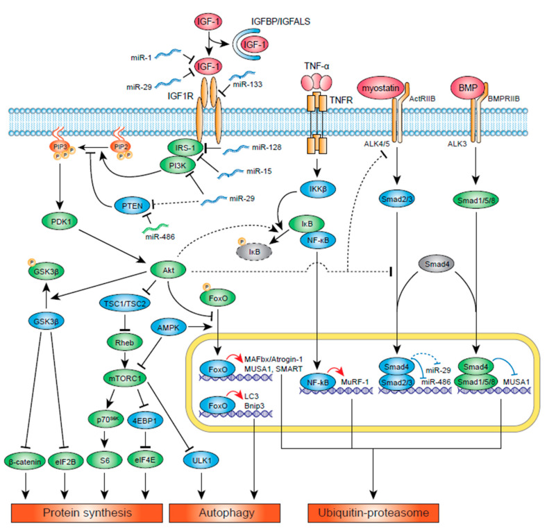
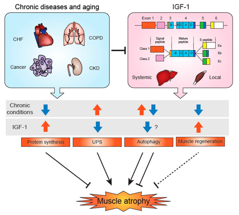
Insulin-like growth factor-1 (IGF-1) is a key growth factor that regulates both anabolic and catabolic pathways in skeletal muscle. IGF-1 increases skeletal muscle protein synthesis via PI3K/Akt/mTOR and PI3K/Akt/GSK3β pathways. PI3K/Akt can also inhibit FoxOs and suppress transcription of E3 ubiquitin ligases that regulate ubiquitin proteasome system (UPS)-mediated protein degradation. Autophagy is likely inhibited by IGF-1 via mTOR and FoxO signaling, although the contribution of autophagy regulation in IGF-1-mediated inhibition of skeletal muscle atrophy remains to be determined. Evidence has suggested that IGF-1/Akt can inhibit muscle atrophy-inducing cytokine and myostatin signaling via inhibition of the NF-κΒ and Smad pathways, respectively. Several miRNAs have been found to regulate IGF-1 signaling in skeletal muscle, and these miRs are likely regulated in different pathological conditions and contribute to the development of muscle atrophy. IGF-1 also potentiates skeletal muscle regeneration via activation of skeletal muscle stem (satellite) cells, which may contribute to muscle hypertrophy and/or inhibit atrophy. Importantly, IGF-1 levels and IGF-1R downstream signaling are suppressed in many chronic disease conditions and likely result in muscle atrophy via the combined effects of altered protein synthesis, UPS activity, autophagy, and muscle regeneration.

Keywords: atrophy; autophagy; cachexia; hypertrophy; insulin-like growth factor-1; muscle regeneration; skeletal muscle.


Conflict of interest statement

The authors declare no conflict of interest.

Figures

Figure 1
Figure 1
IGF-1 signaling pathways. In the figure, the signaling molecules and miRNAs that activate protein synthesis and/or inhibit protein degradation are shown in green, while the ones that inhibit protein synthesis and/or activate protein degradation are shown in blue. The majority of IGF-1 in the body are bound to IGFBP and IGFALS, and its activity is suppressed. Once IGF-1 binds to IGF-1R, IRS-1 and PI3K are recruited and activated. PI3K converts PIP2 to PIP3, which activates PDK1 and Akt. Akt activates protein synthesis via activation of ribosomal protein S6 and the translation initiation factor eIF4E downstream of mTORC1, and activation of β-catenin and eIF2B downstream of GSK3β. Akt can suppress UPS activity via inhibition of FoxO-mediated transcription of E3 ubiquitin ligases MAFbx/Atrogin-1, MUSA1, and SMART. MuRF1 expression is induced by cytokines such as TNF-α via NF-κB pathway. Akt could phosphorylate IκΒ and activate the NF-κΒ pathway, although it has not been shown in skeletal muscle and multiple studies have shown IGF-1 activation does not alter MuRF1 expression. Myostatin and BMP signaling compete against each other for their usage of Smad4. Activation of myostatin inhibits BMP-mediated Smad1/5/8 translocation to the nucleus, thus inhibiting MUSA1-mediated UPS activity. Akt can also downregulate ActRIIB and inhibit ALK4/5 via unknown mechanisms. Although it has not been shown in skeletal muscle, Akt can interact directly with unphosphorylated Smad3 to sequester it outside the nucleus. Several miRNAs have been shown to regulate IGF-1 signaling. miR-486 is inhibited by the myostatin/Smad pathway, resulting in inhibition of IGF-1 signaling via PTEN increase. miR-1 and miR-133 target IGF-1 and IGF-1R, respectively, and their expression is reduced during muscle hypertrophy. IRS-1 could be inhibited by miR-128 and miR-15. LncIRS1 (not shown in the figure), which is upregulated in hypertrophic muscles, can act as sponge for miR-15, resulting in activation of IRS-1. Note that studies have shown conflicting evidence on miR-29′s role in IGF-1 signaling in skeletal muscle, and it may potentiate or inhibit IGF-1 signaling. Pathways that are unclear and/or not shown in skeletal muscle are shown in dotted lines.
Figure 2
Figure 2
IGF-1 and skeletal muscle atrophy in chronic diseases and aging. In various chronic disease conditions, such as congestive heart failure (CHF), cancer, chronic obstructive pulmonary disease (COPD), and chronic kidney disease (CKD), and aging, muscle atrophy develops through various mechanisms: decreased protein synthesis, increased UPS, and lowered muscle regeneration. Depending on the pathophysiological conditions, autophagy could be increased or decreased, and both excessive and defective autophagy could lead to muscle atrophy. IGF-1 is thought to decrease autophagy, but the role of IGF-1 regulation of autophagy in chronic disease-induced muscle atrophy is yet to be determined. IGF-1 stimulates skeletal muscle regeneration via activation of satellite cells. Systemic (circulating) IGF-1 is predominantly produced in the liver, whereas locally produced IGF-1 likely acts in a paracrine or autocrine manner. The first two exons of IGF-1 are mutually exclusive and generate different signal peptides, termed Class 1 (exon 1) and Class 2 (exon 2). The mature IGF-1 peptide is coded in exons 3 and 4 (B, C, A, and D domains). Three types of C-terminus E-peptides are generated by alternative splicing. Ea is from exon 6, Eb is from exon 5, and Ec is from part of exons 5 and 6. Class 2 IGF-1 is mainly expressed in the liver (considered to be the systemic isoform), and Class 1 IGF-1 is mainly expressed in peripheral tissues including skeletal muscle. Both systemic and local IGF-1 levels are decreased in various chronic disease conditions, and the combination of these reductions affect protein synthesis, UPS activity, autophagy, and muscle regeneration and regulate the development of muscle atrophy.

References

    1. Stein A.M., Silva T.M.V., Coelho F.G.M., Arantes F.J., Costa J.L.R., Teodoro E., Santos-Galduroz R.F. Physical exercise, IGF-1 and cognition A systematic review of experimental studies in the elderly. Dement. Neuropsychol. 2018;12:114–122. doi: 10.1590/1980-57642018dn12-020003. - DOI - PMC - PubMed
    1. Majorczyk M., Smolag D. Effect of physical activity on IGF-1 and IGFBP levels in the context of civilization diseases prevention. Rocz. Panstw. Zakl. Hig. 2016;67:105–111. - PubMed
    1. Nindl B.C., Santtila M., Vaara J., Hakkinen K., Kyrolainen H. Circulating IGF-I is associated with fitness and health outcomes in a population of 846 young healthy men. Growth Horm. IGF Res. 2011;21:124–128. doi: 10.1016/j.ghir.2011.03.001. - DOI - PubMed
    1. Maggio M., De Vita F., Lauretani F., Butto V., Bondi G., Cattabiani C., Nouvenne A., Meschi T., Dall’Aglio E., Ceda G.P. IGF-1, the cross road of the nutritional, inflammatory and hormonal pathways to frailty. Nutrients. 2013;5:4184–4205. doi: 10.3390/nu5104184. - DOI - PMC - PubMed
    1. Puche J.E., Castilla-Cortazar I. Human conditions of insulin-like growth factor-I (IGF-I) deficiency. J. Transl. Med. 2012;10:224. doi: 10.1186/1479-5876-10-224. - DOI - PMC - PubMed

Publication types

MeSH terms

Substances

LinkOut - more resources