Review
. 2020 Aug 26;25(17):3891.
doi: 10.3390/molecules25173891.

An Atomic-Level Perspective of HMG-CoA-Reductase: The Target Enzyme to Treat Hypercholesterolemia

Affiliations
Review

An Atomic-Level Perspective of HMG-CoA-Reductase: The Target Enzyme to Treat Hypercholesterolemia

Diana S Gesto et al. Molecules. .

Abstract

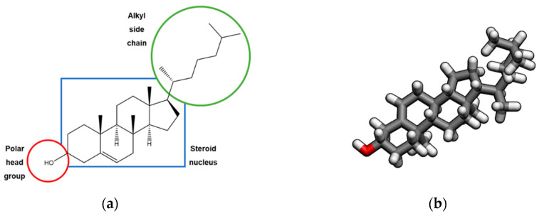
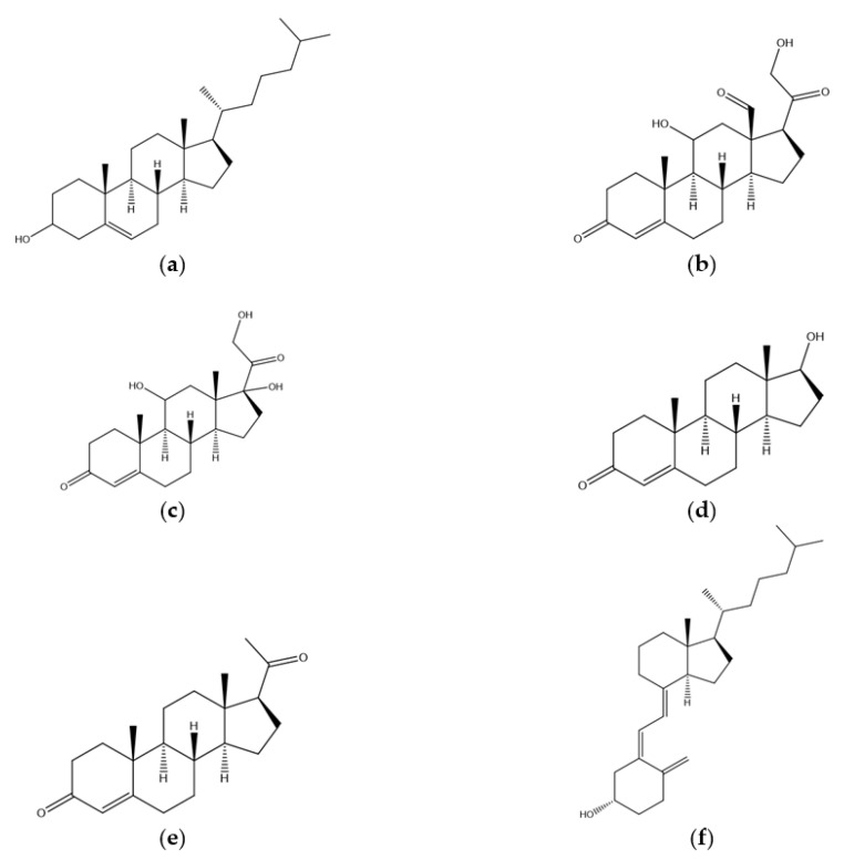
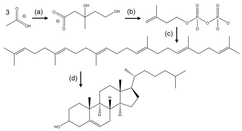
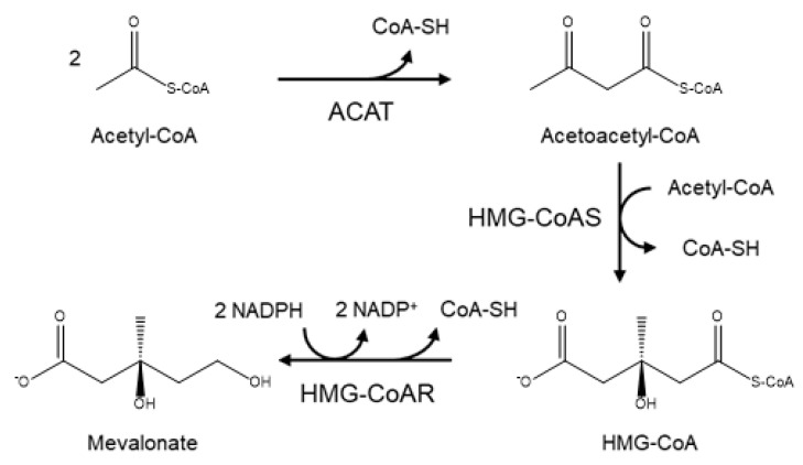
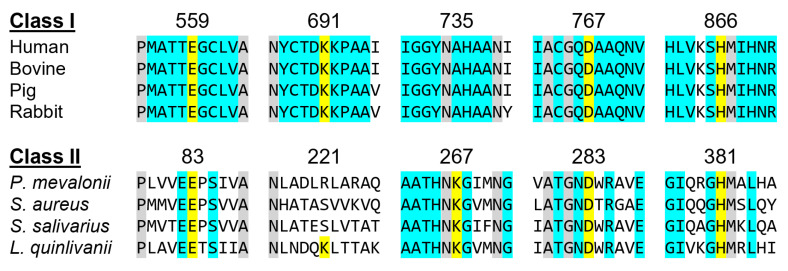
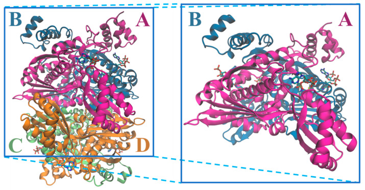
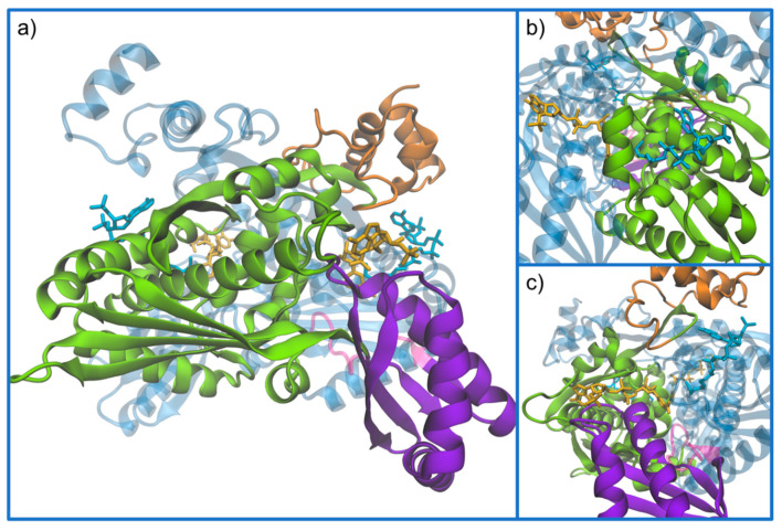
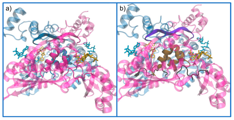
This review provides an updated atomic-level perspective regarding the enzyme 3-hydroxy-3-methylglutaryl coenzyme A reductase (HMG-CoAR), linking the more recent data on this enzyme with a structure/function interpretation. This enzyme catalyzes one of the most important steps in cholesterol biosynthesis and is regarded as one of the most important drug targets in the treatment of hypercholesterolemia. Taking this into consideration, we review in the present article several aspects of this enzyme, including its structure and biochemistry, its catalytic mechanism and different reported and proposed approaches for inhibiting this enzyme, including the commercially available statins or the possibility of using dimerization inhibitors.

Keywords: HMG-CoAR; biochemistry; dimerization inhibitors.; regulation; statins; structure.


Conflict of interest statement

The authors declare no conflict of interest.

Figures

Figure 1
Figure 1
(a) 2D; and (b) 3D structures of the cholesterol molecule.
Figure 2
Figure 2
2D structures of: (a) cholesterol; and some of its precursors: (b) aldosterone; (c) cortisol; (d) testosterone; (e) progesterone; and (f) vitamin-D3.
Figure 3
Figure 3
Summary of cholesterol biosynthesis. (a) Synthesis of mevalonate from acetate; (b) Conversion of mevalonate to activated isoprenes; (c) formation of squalene; (d) conversion of squalene to form the ring steroid nucleus.
Figure 4
Figure 4
Diagram of the mevalonate pathway, the first stage in the biosynthesis of cholesterol.
Figure 5
Figure 5
HMGCRs sequence alignment around the main catalytic residues, which are highlighted in yellow. The numbering is related to the human enzyme for Class I and with the Pseudomonas mevalonii one for Class II. Residues colored in grey are conserved between classes and those colored in light blue are conserved within the same class.
Figure 6
Figure 6
Structure of the tetramer and respective dimer of the human HMGCR, showing both CoA and NADPH (PDB entry 1DQA); chains are colored according with the letters: chain A is in magenta, chain B in blue, chain C in light green and chain D in orange.
Figure 7
Figure 7
(a) Demonstration of the monomer subdomains. The structures follow the representation and color code of the dimer in Figure 6, as chain A and B are colored in magenta and blue (transparent to better see details), respectively. The chain A monomer is subdivided into its subdomains, highlighted in orange is the N-domain; in green the L-domain; in violet the S-domain; and the molecules represented are CoA (in light blue) and NADPH (in yellow). (b,c) Close-up representations of the CoA and NADPH-binding regions, respectively (PDB entry 1DQA).
Figure 8
Figure 8
Demonstration of the dimer interfaces. These structures follow the representation and color code of the dimer in Figure 7, as chain A and B are colored in magenta and blue (transparent to better see details), respectively. (a) The monomer–monomer interactions are detailed by chain; and (b) the characterization of the regions where the interactions are stronger, where the cis-loop, is in black; the β-sheet in violet; and the four α-helix complex in brown (PDB entry 1DQA).
Figure 9
Figure 9
Representation of the conservation score obtained for the catalytic portion of human HMG-CoAR (PDB code 1DQA), using the software ConSurf-DB [55]. It is possible to observe in magenta highly conserved residues, typically with some structural and/or functional importance; the residues with low conservation are in cyan.
Figure 10
Figure 10
Representation of: (A) the active site of HMG-CoAR; (B) the binding sites of the HMG portion of HMG-CoA; and (C) the binding sites of NADP (PDB entry 1DQ9).
Figure 11
Figure 11
Currently accepted catalytic mechanism of HMG-CoAR [56]. The catalytic residues are Lys691, Glu559, Asp767 and His866; chain A is highlighted in pink and chain B is highlighted in blue. Glu559 and His866 are restored in a following step by deprotonation of adjacent water molecules.
Figure 12
Figure 12
Structures of: (a) HMG-CoA; (b) Type I statins; and (c) Type II statins.
Figure 13
Figure 13
Demonstration of the active site residues when a statin (atorvastatin) is bounded (PDB code 1HWK): (a) only atorvastatin is presented in yellow; and (b) HMG-CoA is also bound to more easily compare the inhibitor with the natural substrate (PDB entry 1HWK and 1DQ9). These structures follow the representation and color code of the dimer in Figure 6, as chain A and B are colored in magenta and blue (transparent to better see details), respectively.

References

    1. Windaus A. Über die konstitution des cholesterins und der gallensäuren. Biol. Chem. 1932;213:147–187. doi: 10.1515/bchm2.1932.213.3-4.147. - DOI
    1. Sheppard A.J., O’Dell R.G., Pennington J.A.T. CHOLESTEROL | Properties and determination. In: Caballero B., editor. Encyclopedia of Food Sciences and Nutrition. 2nd ed. Academic Press; Oxford, UK: 2003. pp. 1220–1226.
    1. Nelson D.L., Lehninger A.L., Cox M.M. Lehninger Principles of Biochemistry. W.H. Freeman; New York, NY, USA: 2008.
    1. Arnold D.R., Kwiterovich P.O. CHOLESTEROL | Absorption, Function, and Metabolism. In: Caballero B., editor. Encyclopedia of Food Sciences and Nutrition. 2nd ed. Academic Press; Oxford, UK: 2003. pp. 1226–1237.
    1. Maxfield F.R., van Meer G. Cholesterol, the central lipid of mammalian cells. Curr. Opin. Cell Biol. 2010;22:422–429. doi: 10.1016/j.ceb.2010.05.004. - DOI - PMC - PubMed

MeSH terms

Substances