

Artery of Percheron infarction presenting as nuclear third nerve palsy and transient loss of consciousness: a case report
- PMID: 32859166
- PMCID: PMC7453528
- DOI: 10.1186/s12883-020-01889-9
Artery of Percheron infarction presenting as nuclear third nerve palsy and transient loss of consciousness: a case report
Abstract
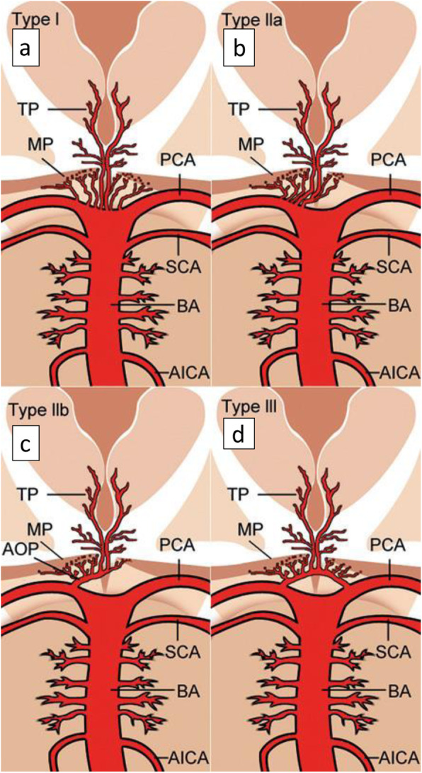
Background: Thalamic blood supply consists of four major vascular territories. Out of them paramedian arteries supply ipsilateral paramedian thalami and occasionally rostral mid brain. Rarely both paramedian arteries arise from a common trunk that arise from P1 segment of one sided posterior cerebral artery (PCA). This is usually due to hypoplastic or absent other P1 and this common trunk is termed Artery of Percheron (AOP). Its prevalence is in the range of 7-11% among the general population and AOP infarcts account in an average of 0.4-0.5% of ischemic strokes. Clinical presentation of AOP infarction is characterized by impaired arousal and memory, language impairment and vertical gaze palsy. It also can present with cerebellar signs, hemi paresis and hemi sensory loss. We herein present a case of AOP infarction presenting as transient loss of consciousness and nuclear third nerve palsy.
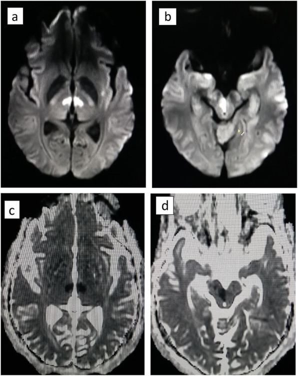
Case presentation: A 51 year old previously healthy male, was brought to us, with a Glasgow coma scale (GCS) of 7/15. GCS improved to 11/15 by the next day, however he had a persisting expressive aphasia. Right sided nuclear third nerve palsy was apparent with the improvement of GCS. He did not have pyramidal or cerebellar signs. Thrombolysis was not offered as the therapeutic window was exceeded by the time of diagnosis. Diagnosis was made using magnetic resonance imaging (MRI) that was done after the initial normal non-contrast computer tomography (NCCT) brain. He was enrolled in stroke rehabilitation. Aspirin and atorvastatin was started for the secondary prevention of stroke. He achieved independency of advanced daily living by 1 month, however could not achieve full recovery to be employed as a taxi driver.
Conclusions: Because of the rarity and varied clinical presentation with altered levels of consciousness, AOP infarcts are easily overlooked as a stroke leading to delayed diagnosis. Timely diagnosis can prevent unnecessary investigations and the patient will be benefitted by early revascularization. As it is seldom reported, case reports remain a valuable source of improving awareness among physicians about this clinical entity.
Keywords: Artery of Percheron (AOP); Case report; Mid brain infarction; Nuclear third nerve palsy; Paramedian thalamic infarction; Thalamic infarction.
Conflict of interest statement
The authors declare that they do not have any competing interests.
Figures

References
Publication types
MeSH terms
LinkOut - more resources
Full Text Sources
Research Materials
