. 2020 Aug 28;20(1):291.
doi: 10.1186/s12876-020-01432-7.

EUS in the diagnosis of pathologically undiagnosed esophageal tuberculosis

Affiliations

EUS in the diagnosis of pathologically undiagnosed esophageal tuberculosis

Rong Zhu et al. BMC Gastroenterol. .

Abstract

Background: Esophageal tuberculosis (ET) is relatively rare, and the diagnosis is challenging. The aim of this study was to evaluate the clinical features of ET and highlight the role of endoscopic ultrasonography (EUS) in the diagnosis of pathologically undiagnosed ET.

Methods: We retrospectively analysed the clinical features, radiological performances, conventional endoscopic appearances, EUS features, treatment and outcomes of pathologically undiagnosed ET between January 2011 and December 2018. All 9 patients failed to be diagnosed by at least two repeated biopsies (such as routine biopsy, multipoint or deep biopsy, and even or EUS-guided fine-needle aspiration (EUS-FNA)).

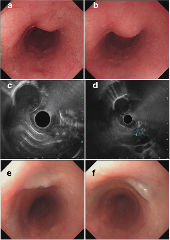
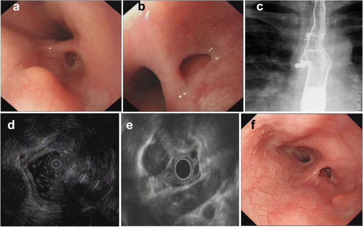
Results: Nine patients (66.7% female) with a mean age of 45 years (range 29-59) complained of retrosternal pain or discomfort, or (and) dysphagia. Esophagoscopy demonstrated protruding lesions in the mucosa with central ulcers or erosion in five patients, submucosal bulges with smooth surfaces in one patient, submucosal bulges with diverticula in one patient, ulcers with suspicious fistula formation in one patient, and multiple ulcers in one patient. None of the patients received confirmed histopathological or bacteriological diagnoses by repeated biopsies. However, they were first suspected to have ET based on EUS examination. Because EUS found some characteristic ultrasonographic changes, which were very helpful for the diagnosis of ET when combined with clinical manifestations, the patients subsequently received diagnostic antituberculosis therapy. Finally, the patients recovered or improved with follow-up times ranging from 3 to 10 months.

Conclusions: EUS could help in the diagnosis of ET on basis of EUS features like poorly defined esophageal wall structure, enlarged paraesophageal or mediastinal lymph nodes, hypoechoic lesions of esophageal wall that are linked to the enlarged paraesophageal lymph nodes. However all attempts should be made to obtain histological or microbiological diagnosis.

Keywords: Dysphagia; Endoscopic ultrasonography (EUS); Esophageal tuberculosis (ET); Retrosternal pain.


Conflict of interest statement

The authors declare that they have no competing interests.

Figures

Fig. 1
Fig. 1
Esophagoscopic findings and EUS features of pathologically undiagnosed ET (cases 1–3). 1–3, case 1–3. a (far view) and b (near view) Esophagoscopy: a protruding lesion in the mucosa with a central ulcer. c and d, EUS: thickening, poorly defined esophageal wall structure, with one or more enlarged paraesophageal lymph nodes. e, repeat esophagoscopy after treatment: a white mucosal depression with scar formation
Fig. 2
Fig. 2
Esophagoscopic findings and EUS features of pathologically undiagnosed ET (cases 4 and 5). 1, case 4; 2, case 5. a (far view) and b (near view) Esophagoscopy: a protruding lesion in the mucosa with central erosion. c and d, EUS: thickening, poorly defined esophageal wall structure, with (case 4) or without (case 5) enlarged paraesophageal lymph nodes. e, repeat esophagoscopy after treatment: a white mucosal depression with scar formation (case 4) and the protruding lesion in the mucosa became smaller with disappearance of erosion (case 5)
Fig. 3
Fig. 3
Esophagoscopic findings and EUS features of pathologically undiagnosed ET (case 6). a (far view) and b (near view) Esophagoscopy: a submucosal bulge with smooth surface. c and d, EUS: thickening, poorly defined esophageal wall structure, linked to an enlarged paraesophageal lymph node, with scattered calcifications. e and f, repeat esophagoscopy after treatment: a white mucosal depression with scar formation
Fig. 4
Fig. 4
Esophagoscopic findings and EUS features of pathologically undiagnosed ET (case 7). a (far view) and b (near view) Esophagoscopy: a submucosal bulge and diverticulum. c, Esophagography: a diverticulum formation. d and e, EUS: thickening, poorly defined esophageal wall structure, with enlarged paraesophageal lymph nodes. f, repeat esophagoscopy after treatment: the submucosal bulge became smaller
Fig. 5
Fig. 5
Esophagoscopic findings and EUS features of pathologically undiagnosed ET (case 8). a (far view) and b (near view) Esophagoscopy: an ulcer with suspicious fistula formation. c, chest CT: a small amount of bubbles and fluid in the mediastinum around the esophagus. d and e, EUS: thickening, poorly defined esophageal wall structure, with one or more enlarged paraesophageal lymph nodes. f, repeat esophagoscopy after treatment: a white mucosal depression with scar formation
Fig. 6
Fig. 6
Esophagoscopic findings and EUS features of pathologically undiagnosed ET (case 9). a (far view) and b (near view) Esophagoscopy: multiple ulcers. c and d, EUS: Thickening, poorly defined esophageal wall structure, with an enlarged paraesophageal lymph node. e, repeat esophagoscopy 2 months after treatment: the ulcers improved. f, repeat esophagoscopy 4 months after treatment: white mucosal depressions with scar formation

References

    1. Lockard LB. Oesophageal tuberculosis: a critical review. Laryngoscope. 1913;23:561–584. doi: 10.1288/00005537-191305000-00001. - DOI
    1. Carr DT, Spain DM. Tuberculosis in a carcinoma of the esophagus. Am Rev Tubercul. 1942;46:346–349.
    1. Jain SK, Jain S, Jain M, et al. Esophageal tuberculosis: is it so rare? Report of 12 cases and review of the literature. Am J Gastroenterol. 2002;97(2):287–294. doi: 10.1111/j.1572-0241.2002.05456.x. - DOI - PubMed
    1. Rana SS, Mandavdhare H, Sharma V, et al. Successful closure of chronic, nonhealing tubercular esophagobronchial fistula with an over-the-scope clip. J Digestive Endoscopy. 2017;8(1):33–35. doi: 10.4103/0976-5042.202820. - DOI
    1. Dahale AS, Kumar A, Srivastava S, et al. Esophageal tuberculosis: uncommon of common. JGH Open. 2018;2(2):34–38. doi: 10.1002/jgh3.12043. - DOI - PMC - PubMed