. 2021 Jan;126(1):245-255.
doi: 10.1016/j.bja.2020.07.022. Epub 2020 Aug 25.

The T-type calcium channel isoform Cav3.1 is a target for the hypnotic effect of the anaesthetic neurosteroid (3β,5β,17β)-3-hydroxyandrostane-17-carbonitrile

Affiliations

The T-type calcium channel isoform Cav3.1 is a target for the hypnotic effect of the anaesthetic neurosteroid (3β,5β,17β)-3-hydroxyandrostane-17-carbonitrile

Tamara Timic Stamenic et al. Br J Anaesth. 2021 Jan.

Abstract

Background: The mechanisms underlying the role of T-type calcium channels (T-channels) in thalamocortical excitability and oscillations in vivo during neurosteroid-induced hypnosis are largely unknown.

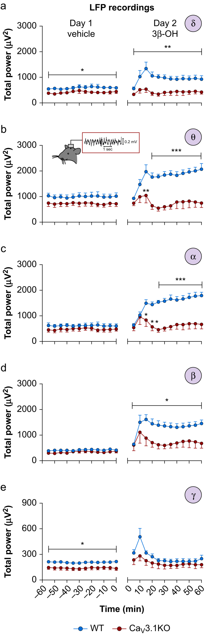
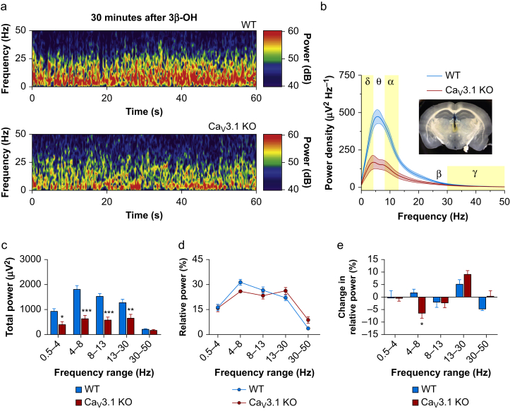
Methods: We used patch-clamp electrophysiological recordings from acute brain slices ex vivo, recordings of local field potentials (LFPs) from the central medial thalamic nucleus in vivo, and wild-type (WT) and Cav3.1 knock-out mice to investigate the molecular mechanisms of hypnosis induced by the neurosteroid analogue (3β,5β,17β)-3-hydroxyandrostane-17-carbonitrile (3β-OH).

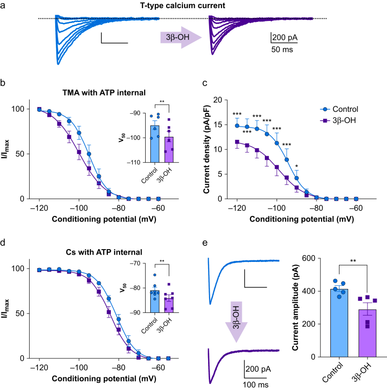
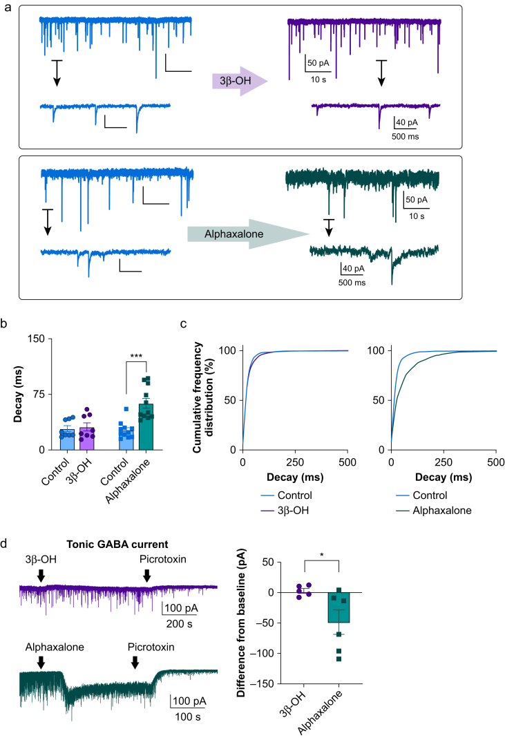
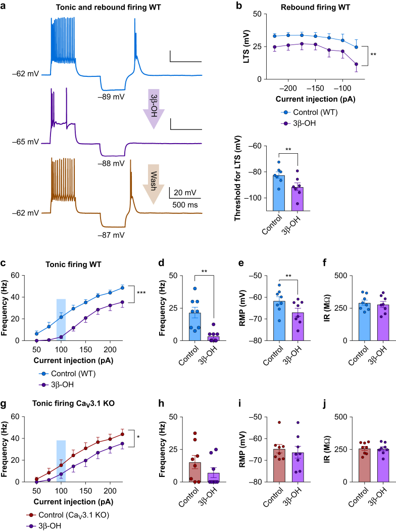
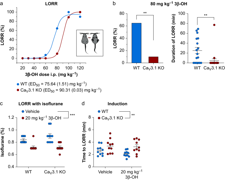
Results: Patch-clamp recordings showed that 3β-OH inhibited isolated T-currents but had no effect on phasic or tonic γ-aminobutyric acid A currents. Also in acute brain slices, 3β-OH inhibited the spike firing mode more profoundly in WT than in Cav3.1 knockout mice. Furthermore, 3β-OH significantly hyperpolarised neurones, reduced the amplitudes of low threshold spikes, and diminished rebound burst firing only in WT mice. We found that 80 mg kg-1 i.p. injections of 3β-OH induced hypnosis in >60% of WT mice but failed to induce hypnosis in the majority of mutant mice. A subhypnotic dose of 3β-OH (20 mg kg-1 i.p.) accelerated induction of hypnosis by isoflurane only in WT mice, but had similar effects on the maintenance of isoflurane-induced hypnosis in both WT and Cav3.1 knockout mice. In vivo recordings of LFPs showed that a hypnotic dose of 3β-OH increased δ, θ, α, and β oscillations in WT mice in comparison with Cav3.1 knock-out mice.

Conclusions: The Cav3.1 T-channel isoform is critical for diminished thalamocortical excitability and oscillations that underlie neurosteroid-induced hypnosis.

Keywords: calcium channel; electrophysiology; hypnosis; mechanisms of anaesthesia; neurosteroid; thalamus.


Figures

Figure 1
Figure 1
Inhibition of T-currents by 3β-OH in CeM neurones. (a) Original traces of T currents in a representative CeM neurone in control conditions recorded using a double-pulse protocol with 3.6 s long prepulses to variable voltages (from –115 to –80 mV in 5 mV increment; blue); traces from the same cell using the identical voltage protocol during an apparent steady-state inhibition of T-current by 3 μM 3β-OH (purple) recorded with TMA with ATP internal solution. (b) Average normalised steady-state inactivation (I/Imax) curves in control conditions and after application of 3β-OH in the same cells. 3β-OH induced a hyperpolarising shift in V50 of 4.6 mV; the average V50 value for steady-state inactivation was –94.9 (1.8) mV in control conditions (blue; six cells, three animals). After 3β-OH perfusion of the same cells, the V50 value was –99.5 (2.1) mV (purple). In the inset of panel (b), the V50 was significantly different after 3β-OH perfusion (two tailed paired t-test: t(5)=4.08, P<0.01). (c) Average current density calculated from the steady-state inactivation protocol. Application of 3β-OH (purple line and data points; six cells, three animals) decreased current density when compared with control conditions (blue line and data points). Data were analysed with two-way RM analysis of variance (anova): interaction F(12,60)=13.91, P<0.001; voltage F(12.60)=46.66, P<0.001; 3β-OH F(1,5)=15.81, P=0.011; Sidak's post hoc presented in figure). (d) Average normalised steady-state inactivation (I/Imax) curves in control conditions and after application of 3β-OH in the same cells recorded with Cs with ATP internal solution. 3β-OH induced a hyperpolarising shift in V50 of 3.2 mV because the average V50 value for steady-state inactivation was –80.9 (1.2) mV under control conditions (blue; seven cells, four animals), and after 3β-OH perfusion in the same cells, the V50 value was –84.1 (1.3) mV (purple). In the inset of panel (d), the V50 was significantly different after 3β-OH perfusion (two tailed paired t-test: t(6)=3.92, P=0.008). (e) Averaged representative traces recorded under control conditions (blue) and after application of 3β-OH (purple) using a protocol Vt=−50 mV, Vh=−90 mV, 3β-OH decreased T-current amplitudes from 416.9 (17.5) pA to 291.9 (38.2) pA (two-tailed paired t-test: t(4)=4.67, P<0.01; five cells, three animals) ∗P<0.05, ∗∗P<0.01, ∗∗∗P<0.001. 3β-OH, (3β,5β,17β)-3-hydroxyandrostane-17-carbonitrile; ATP, adenosine triphosphate; CeM, central medial nucleus; RM, repeated measure; TMA, tetramethylammonium.
Figure 2
Figure 2
Potentiation of miniature inhibitory postsynaptic currents by alphaxalone but not by 3β-OH. (a) Representative traces from CeM neurones before and after perfusion with 3β-OH (top panel) or alphaxalone (bottom panel). (b) Decay tau was increased only after perfusion of alphaxalone but not 3β-OH; alphaxalone increased decay tau from 26.7 (3.4) to 62.9 (6.6) ms (two-tailed paired t-test: t(10)=6.82, P<0.001). (c) Left panel shows that cumulative frequency distribution for decay tau confirms no change after application of 3β-OH (control 1479 events, 3β-OH 1497 events); right panel shows longer decay taus after alphaxalone perfusion (1523 events for alphaxalone, 2134 events for the control). (d) Representative traces showing tonic GABA current after 3 μM 3β-OH (purple trace) or 3 μM alphaxalone (green trace) perfusion. (b) Difference from baseline showed a minimal effect of 3β-OH on tonic current in comparison with alphaxalone (3β-OH five cells, alphaxalone six cells; unpaired two-tailed t-test: t(9)=2.31, P=0.046). ∗P<0.05, ∗∗∗P<0.001. 3β-OH, (3β,5β,17β)-3-hydroxyandrostane-17-carbonitrile; CeM, central medial nucleus; GABA, γ-aminobutyric acid A.
Figure 3
Figure 3
3β-OH influence on the excitability of CeM neurons. (a) Representative traces from a WT CeM neurone before application of 3β-OH (blue), after application of 3 μM 3β-OH (purple) and after wash (orange trace) with active membrane responses to a depolarising (125 pA) and hyperpolarising (–75 pA) current injection. (b) Top: averaged LTS amplitude was reduced by application of 3β-OH across all hyperpolarising current pulses from –50 to –225 pA in WT mice (eight cells, four animals; two-way RM analysis of variance [anova]: interaction F(6,42)=0.41, P=0.865; current injection F(6,42)=5.44, P<0.001; 3β-OH F(1,7)=22.63, P=0.002). Bottom: bar graph shows 3β-OH increased the threshold for the occurrence of LTS (seven cells, four animals; paired two-tailed t-test t(6)=4.07, P=0.007). (c) 3β-OH reduced tonic action potential firing frequency across all current pulses in WT mice (eight cells, four animals; from 50 to 225 pA, two-way RM anova: interaction F(7,49)=1.66, P=0.140; current injection F(7,49)=132.70, P<0.001; 3β-OH effect F(1,7)=29.52, P=0.001). (d) 3β-OH decreased tonic frequency at 100 pA current injection in WT mice (eight cells, four animals; paired two-tailed t-test: t(7)=4.80, P=0.002). (e) 3β-OH hyperpolarised RMP in WT mice (eight cells, four animals; paired two-tailed t-test: t(7)=4.00, P=0.005). (f) 3β-OH did not affect IR measured with a hyperpolarising current injection of 100 pA in WT mice. (g) 3β-OH diminished tonic action potential firing frequency in Cav3.1 KO mice (eight cells, three animals; from 50 to 225 pA, two-way RM anova: interaction F(7,49)=1.23, P=0.302; current injection F(7,49)=63.29, P<0.001; 3β-OH effect F(1,7)=8.26, P=0.024). (h) 3β-OH did not significantly affect tonic firing frequency at 100 pA current injection in Cav3.1 KO mice. (i) 3β-OH did not significantly affect RMP in Cav3.1 KO mice. (j) 3β-OH did not significantly affect IR measured with a hyperpolarising current injection of 100 pA in Cav3.1 KO mice. ∗P<0.05, ∗∗P<0.01, ∗∗∗P<0.001. 3β-OH, (3β,5β,17β)-3-hydroxyandrostane-17-carbonitrile; CeM, central medial nucleus; GABA, γ-aminobutyric acid A; KO, knock-out; LTS, low threshold spikes; RM, definition; IR, input resistance; RMP, resting membrane potential; WT, wild-type.
Figure 4
Figure 4
Effect of 3β-OH on anaesthetic endpoints. (a) Dose–response curve for loss of righting reflex (LORR) in WT and Cav3.1 KO mice with ED50 indicated on the top of the panel. (b) Left, percentage of animals that lost righting reflex under 80 mg kg−1 i.p. 3β-OH in WT (17 animals) and Cav3.1 KO (20 animals) cohorts (80 mg kg−1: χ2 = 9.79, P=0.002). (b) Right: duration of LORR under 80 mg kg−1 i.p. 3β-OH in WT (17 animals) and Cav3.1 KO (20 animals) mice (unpaired two-tailed t-test: t(35)=2.94, P=0.006). (c) 20 mg kg−1 3β-OH lowered the percent of isoflurane needed for LORR in WT and Cav3.1 KO mice (10 mice in each group, two-way RM analysis of variance [anova]: interaction F(1,18)=1.67, P=0.210; mutation effect F(1,18)=1.35, P=0.260; 3β-OH effect F(1,18)=42.19, P<0.001). (d) WT animals pretreated with 3β-OH needed significantly less time for induction with isoflurane compared with mutant animals (10 mice in each group, two-way RM anova: interaction F(1,18)=0.71, P=0.410; mutation effect F(1,18)=9.05, P=0.007; 3β-OH effect F(1,18)=1.59, P=0.223). ∗∗P<0.01, ∗∗∗P<0.001. 3β-OH, (3β,5β,17β)-3-hydroxyandrostane-17-carbonitrile; KO, knock-out; WT, wild-type; ED50, median effective dose; RM, definition.
Figure 5
Figure 5
Oscillations in CeM neurones after injections of 3β-OH. (a) Total δ power after injections of the vehicle 2-hydroxypropyl-β-cyclodextrin (Day 1) and 3β-OH (Day 2) in WT and Cav3.1 KO mice. Mutant mice had less δ power on Day 1 (two-way RM analysis of variance [anova]: interaction F(11,121)=0.27, P=0.990; time F(11,121)=1.15, P=0.330; mutation effect F(1,11)=5.93, P=0.033). 3β-OH enhanced δ power in WT animals (two-way RM anova: interaction F(11,121)=1.51, P=0.137; time F(11,121)=3.62, P<0.001; mutation effect F(1,11)=11.99, P=0.005). (b) Total θ power after vehicle (Day 1) or 3β-OH (Day 2) in WT and Cav3.1 KO mice. 3β-OH enhanced θ power in WT animals (two-way RM anova: interaction F(11,121)=4.07, P<0.001; time F(11,121)=4.03, P<0.001; mutation effect F(1,11)=26.60, P<0.001; Sidak's post hoc presented in figure). (c) Total α power for vehicle (Day 1) or 3β-OH (Day 2) in WT and Cav3.1 KO mice. 3β-OH enhanced α power in WT animals (two-way RM anova: interaction F(11,121)=5.87, P<0.001; time F(11,121)=5.10, P<0.001; mutation effect F(1,11)=27.74, P<0.001; Sidak's post hoc presented in figure). (d) Total β power for vehicle (Day 1) or 3β-OH (Day 2) in WT and Cav3.1 KO mice. 3β-OH enhanced β power in WT animals (two-way RM anova: interaction F(11,121)=1.16, P=0.324; time F(11,121)=2.82, P=0.003; mutation effect F(1,11)=8.62, P=0.013; Sidak's post hoc presented in figure). (e) Total γ power for vehicle (Day 1) or 3β-OH (Day 2) in WT and Cav3.1 KO mice. Mutant mice had less γ power on Day 1 (two-way RM anova: interaction F(11,121)=0.53, P=0.879; time F(11,121)=1.77, P=0.067; mutation effect F(1,11)=6.73, P=0.025). 3β-OH did not statistically change γ power between WT and Cav3.1 KO animals. WT, seven animals, Cav3.1 KO, six animals; ∗P<0.05, ∗∗P<0.01, ∗∗∗P<0.001. 3β-OH, (3β,5β,17β)-3-hydroxyandrostane-17-carbonitrile; CeM, central medial nucleus; KO, knock-out; LFP, local field potentials; WT, wild-type.
Figure 6
Figure 6
Oscillatory differences in CeM between WT and Cav3.1 KO mice after 3β-OH. (a) Representative spectrograms from the CeM at 30 min after i.p. injections of 3β-OH recorded from WT (upper image) and Cav3.1 KO mice (lower image). (b) Power density with electrode confirmation (inset in figure). Power density revealed decreased low frequency oscillations in Cav3.1 KO mice under 3β-OH. (c) Total power revealed less power in δ, θ, α, and β frequency range in Cav3.1 KO mice (two-way RM analysis of variance [anova]: interaction F(4,44)=10.76, P<0.001; oscillations F(4,44)=37.80, P<0.001; mutation effect F(1,11)=32.50, P<0.001; Sidak's post hoc presented in figure). (d) Analysis of relative power after neurosteroid injection did not find significant differences between WT and Cav3.1 KO mice. (e) Change in relative power (relative power under 3β-OH minus relative power during baseline in CeM) revealed differences between WT and Cav3.1 KO mice in the θ frequency range (two-way RM anova: interaction F(4,44)=3.30, P=0.019; oscillations F(4,44)=8.08, P<0.001; mutation effect F(1,11)=0.31, P=0.591; Sidak's post hoc presented in figure). WT, seven animals; Cav3.1 KO, six animals; ∗P<0.05, ∗∗P<0.01, ∗∗∗P<0.001. 3β-OH, (3β,5β,17β)-3-hydroxyandrostane-17-carbonitrile; CeM, central medial nucleus; KO, knock-out; RM, repeated measure; WT, wild-type.

Comment in

References

    1. Atluri N., Joksimovic S.M., Oklopcic A. A neurosteroid analogue with T-type calcium channel blocking properties is an effective hypnotic, but is not harmful to neonatal rat brain. Br J Anaesth. 2018;120:768–778. - PMC - PubMed
    1. Rupprecht R. Neuroactive steroids: mechanisms of action and neuropsychopharmacological properties. Psychoneuroendocrinology. 2003;28:139–168. - PubMed
    1. Stell B.M., Brickley S.G., Tang C.Y., Farrant M., Mody I. Neuroactive steroids reduce neuronal excitability by selectively enhancing tonic inhibition mediated by subunit-containing GABAA receptors. Proc Natl Acad Sci. 2003;100:14439–14444. - PMC - PubMed
    1. Lambert J.J., Belelli D., Hill-Venning C., Peters J.A. Neurosteroids and GABAA receptor function. Trends Pharmacol Sci. 1995;16:295–303. - PubMed
    1. Tuem K.B., Atey T.M. Neuroactive steroids: receptor interactions and responses. Front Neurol. 2017;8:1–10. - PMC - PubMed