. 2020 Dec:551:84-92.
doi: 10.1016/j.virol.2020.05.014. Epub 2020 Jun 22.

Bat mammalian orthoreoviruses cause severe pneumonia in mice

Affiliations

Bat mammalian orthoreoviruses cause severe pneumonia in mice

Ren-Di Jiang et al. Virology. 2020 Dec.

Abstract

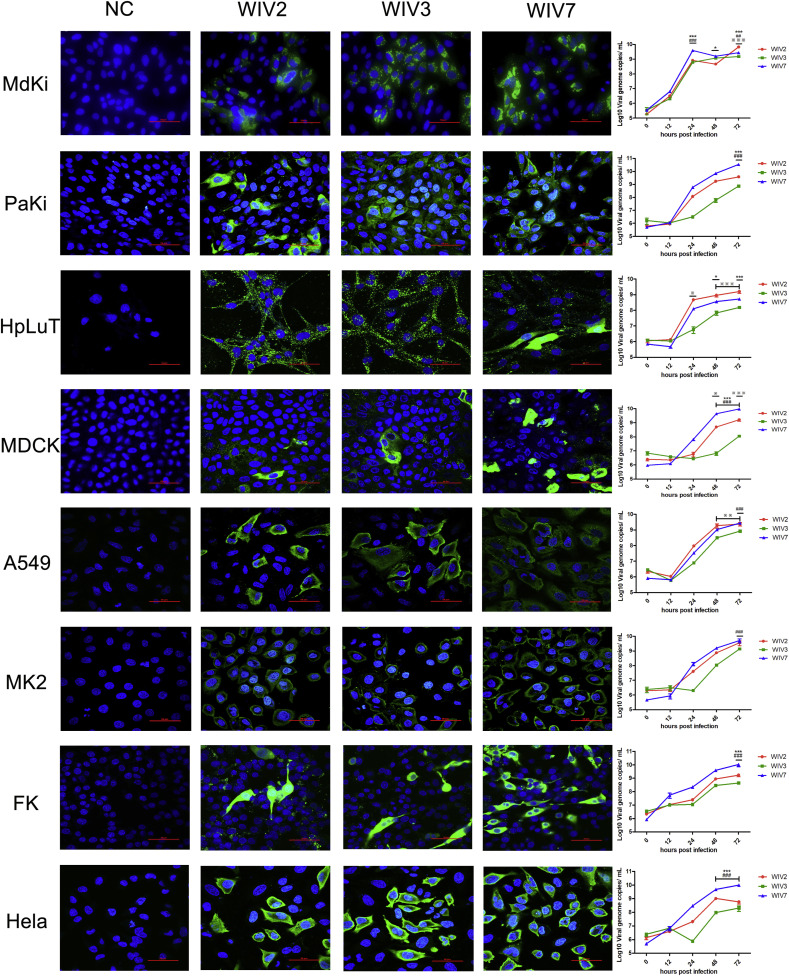
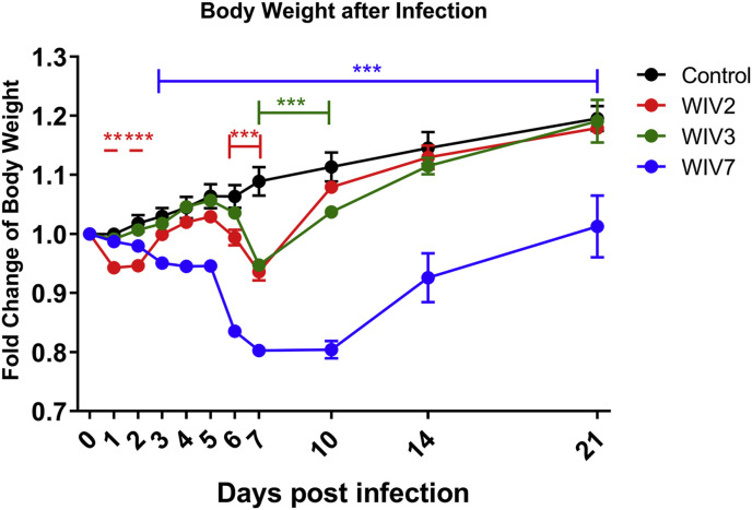
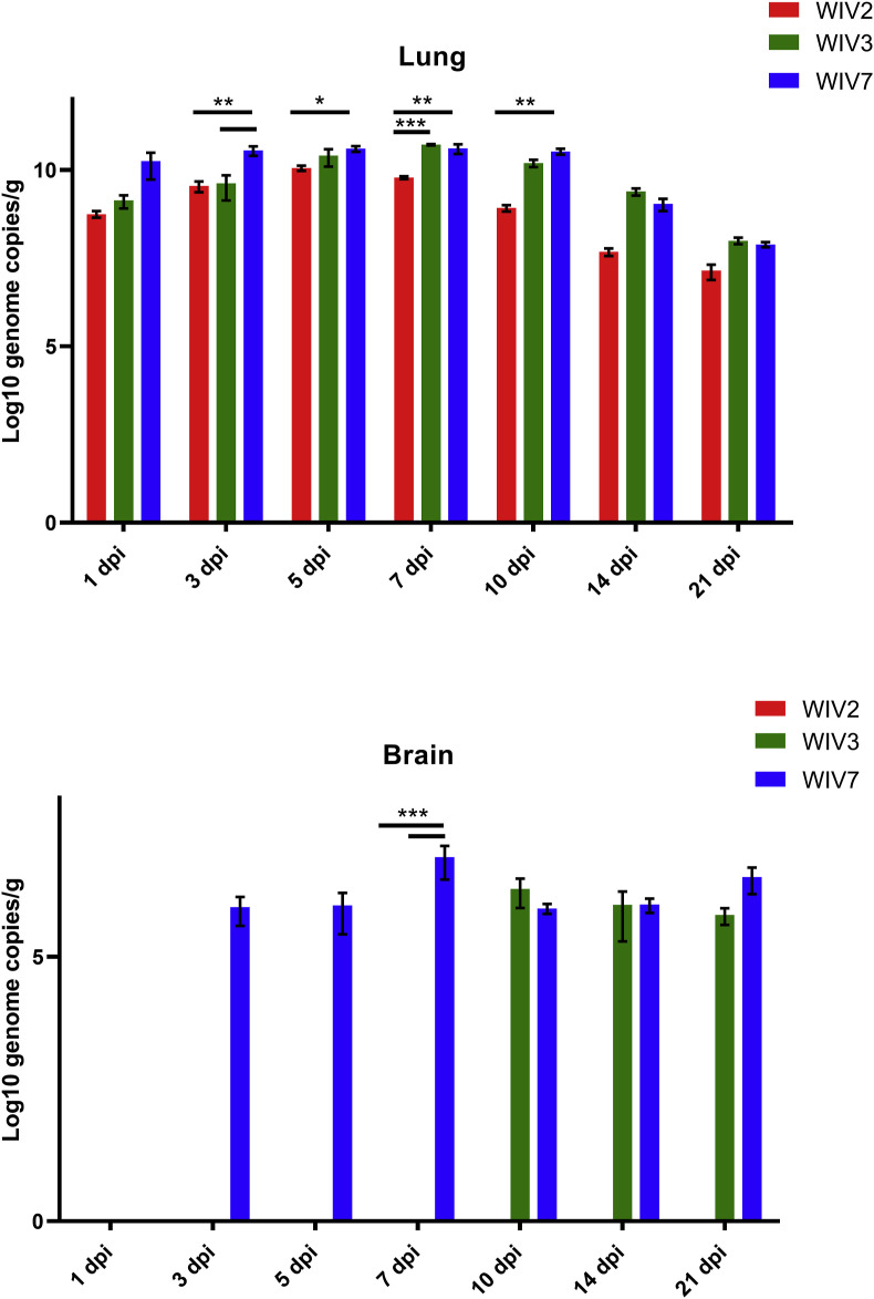
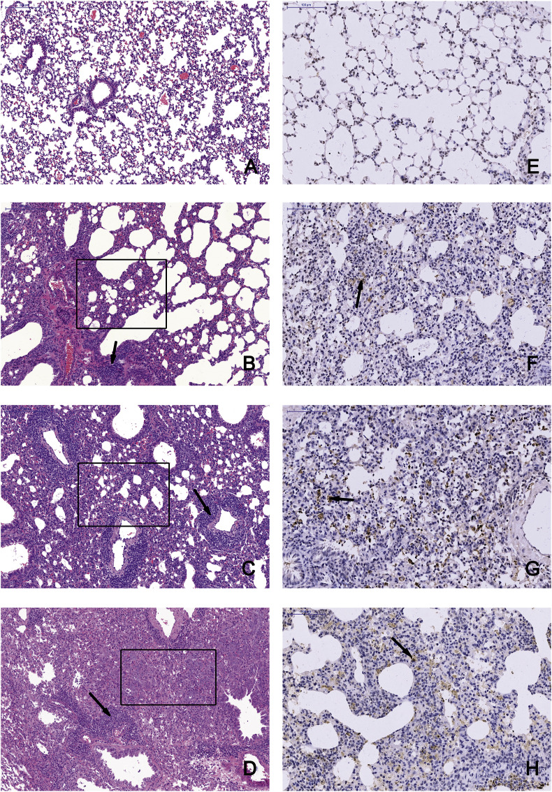
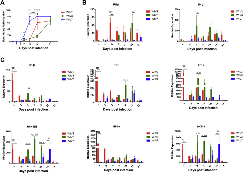
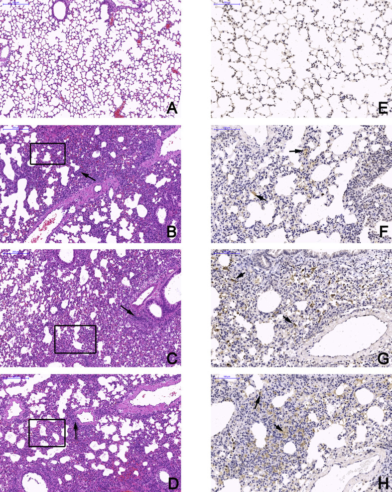
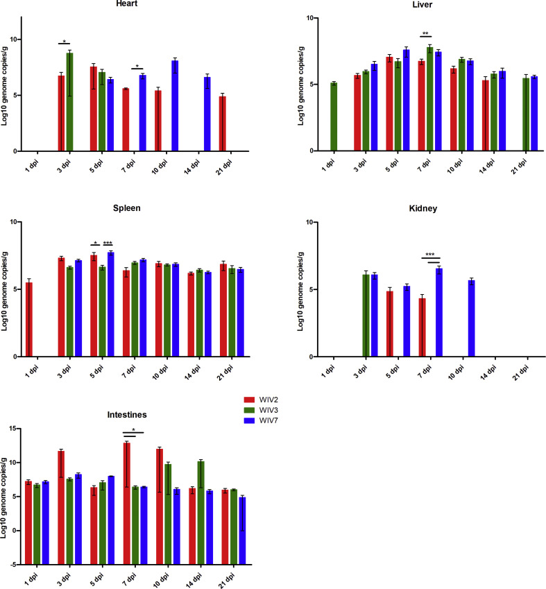
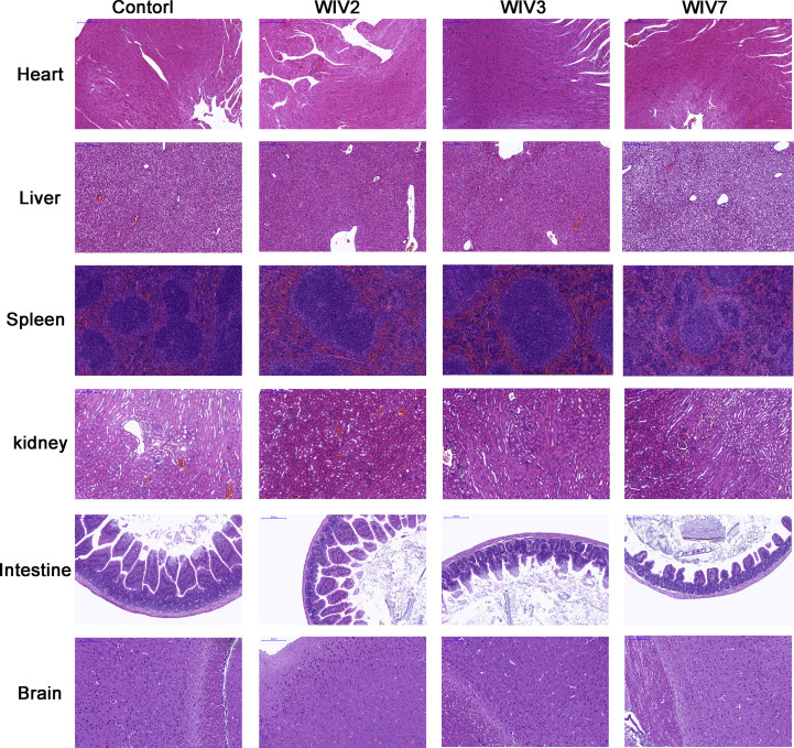
Mammalian orthoreovirus (MRV) infections are ubiquitous in mammals. Increasing evidence suggests that some MRVs can cause severe respiratory disease and encephalitis in humans and other animals. Previously, we isolated six bat MRV strains. However, the pathogenicity of these bat viruses remains unclear. In this study, we investigated the host range and pathogenicity of 3 bat MRV strains (WIV2, 3 and 7) which represent three serotypes. Our results showed that all of them can infect cell lines from different mammalian species and displayed different replication efficiency. The BALB/c mice infected by bat MRVs showed clinical symptoms with systematic infection especially in lung and intestines. Obvious tissue damage were found in all infected lungs. One of the strains, WIV7, showed higher replication efficiency in vitro and vivo and more severe pathogenesis in mice. Our results provide new evidence showing potential pathogenicity of bat MRVs in animals and probable risk in humans.

Keywords: Bat; Mammalian orthoreovirus; Pathogenicity; Pneumonia.


Figures

Fig. 1
Fig. 1
Bat MRVs have wide cell tropism. MdKi, PaKi, HpLuT, MDCK, A549, LLC-MK2, FK and Hela cells were infected with bat MRV WIV2, WIV3 and WIV7 (MOI = 1). Immunofluorescence assay (Panel 1–4) was performed at 24 hpi. Bat MRV antigens were stained with rabbit anti-WIV3 polyclonal antibody as the primary antibody followed by FITC-labelled mouse anti-rabbit IgG and coloured in green. Cell nuclei were stained with DAPI and shown in blue. Scale bar is 50 μm. Multi-cycle supernatants were taken at 0, 12, 24, 48 and 72 hpi. Viral loads were determined by RT-qPCR (panel 5). Error bar indicates the standard error. * represent WIV-2 group compared with WIV-7 group, # represent WIV-3 group compared with WIV-7 group, ※ represent WIV-2 group compared with WIV-3 group. *P < 0.05, **P < 0.01, ***P < 0.001.
Fig. 2
Fig. 2
Body weight changes after viral infection. Four-week-old female BALB/c mice were infected with 105 TCID50 bat MRV WIV2, WIV3 or WIV7 via the internasal route. Body weights were measured at 0, 1, 2, 3, 4, 5, 6, 7, 10, 14 and 21 days post-infection. Error bar indicates the standard error. **P < 0.01, ***P < 0.001, compared with control group.
Fig. 3
Fig. 3
Viral RNA load in lung and brain tissues after bat MRV infection. Lung and brain from infected BALB/c mice at 1, 3, 5, 7, 10, 14 and 21 dpi were detected for viral RNA load. WIV2, WIV3 and WIV7 show different infection efficiency in BALB/c mice. N = 3 in each time point. Error bar indicates the standard error. Black underline represent the comparison between indicated groups, *P < 0.05, **P < 0.01, ***P < 0.001.
Fig. 4
Fig. 4
Bat MRVs infection cause severe pneumonia in BALB/c mice. Left lung lobe of BALB/c mice at 14 dpi were subjected to pathological exam by H&E staining and IHC assay, images were taken from the base of lungs. A and E, mock-infected mice examined by H&E staining and IHC, respectively. B-D, mice infected by WIV2, WIV3 and WIV7, respectively, was examined by H&E staining. The tested tissues showed severe pneumonia with distinct alveolar thickening, reduction of bronchioles and alveoli (black box) and lymphocytic infiltration (black arrow). Scale bar is 200 μm. For IHC assay, rabbit anti-WIV3 polyclonal antibody was used as the primary antibody and HRP-labelled goat anti-rabbit IgG was used as the secondary antibody. The positive area is brownish yellow and can be observed in WIV2- (F), WIV3- (G) and WIV7 (H)-infected lungs. Scale bar is 100 μm.
Fig. 5
Fig. 5
Bat MRVs induced different patterns of humoral immune response and proinflammatory gene production in BALB/c mice. Infected and mock-infected mice were euthanised at several time points, serum was separated from blood and lungs were collected. Anti-bat MRV neutralisation antibody titres were determined (A). Each serum was measured in triplicate. Titres were expressed as the reciprocal of the final serum dilution required to neutralise all inoculated wells. Total lung RNA was analysed for mRNA expression by RT-qPCR. The gene transcription levels of IFN-β, IFN-γ (B) and proinflammatory cytokines/chemokines (C) were assessed. All data were normalised to 18S rRNA. Relative expression was expressed as the relative fold increase over mock-infected mice. Error bar indicates the standard error. * represent WIV-2 group compared with WIV-7 group, # represent WIV-3 group compared with WIV-7 group, ※ represent WIV-2 group compared with WIV-3 group (A). Black underline represent the comparison between indicated groups (B and C), *P < 0.05, **P < 0.01, ***P < 0.001.
figs1
figs1
figs2
figs2
figs3
figs3

References

    1. Akdis M., Aab A., Altunbulakli C., Azkur K., Costa R.A., Crameri R., Duan S., Eiwegger T., Eljaszewicz A., Ferstl R., Frei R., Garbani M., Globinska A., Hess L., Huitema C., Kubo T., Komlosi Z., Konieczna P., Kovacs N., Kucuksezer U.C., Meyer N., Morita H., Olzhausen J., O'Mahony L., Pezer M., Prati M., Rebane A., Rhyner C., Rinaldi A., Sokolowska M., Stanic B., Sugita K., Treis A., van de Veen W., Wanke K., Wawrzyniak M., Wawrzyniak P., Wirz O.F., Zakzuk J.S., Akdis C.A. Interleukins (from IL-1 to IL-38), interferons, transforming growth factor beta, and TNF-alpha: receptors, functions, and roles in diseases. J. Allergy Clin. Immunol. 2016;138:984–1010. - PubMed
    1. Anafu A.A., Bowen C.H., Chin C.R., Brass A.L., Holm G.H. Interferon-inducible transmembrane protein 3 (IFITM3) restricts reovirus cell entry. J. Biol. Chem. 2013;288:17261–17271. - PMC - PubMed
    1. Antar A.A., Konopka J.L., Campbell J.A., Henry R.A., Perdigoto A.L., Carter B.D., Pozzi A., Abel T.W., Dermody T.S. Junctional adhesion molecule-A is required for hematogenous dissemination of reovirus. Cell Host Microbe. 2009;5:59–71. - PMC - PubMed
    1. Attoui H., Biagini P., Stirling J., Mertens P.P., Cantaloube J.F., Meyer A., de Micco P., de Lamballerie X. Sequence characterization of Ndelle virus genome segments 1, 5, 7, 8, and 10: evidence for reassignment to the genus Orthoreovirus, family Reoviridae. Biochem. Biophys. Res. Commun. 2001;287:583–588. - PubMed
    1. Berard A.R., Cortens J.P., Krokhin O., Wilkins J.A., Severini A., Coombs K.M. Quantification of the host response proteome after mammalian reovirus T1L infection. PLoS One. 2012;7 - PMC - PubMed

Publication types