

Tracheal swab from front of neck airway for SARS-CoV-2; a bronchial foreign body
- PMID: 32859619
- PMCID: PMC10577772
- DOI: 10.1136/bcr-2020-237787
Tracheal swab from front of neck airway for SARS-CoV-2; a bronchial foreign body
Abstract
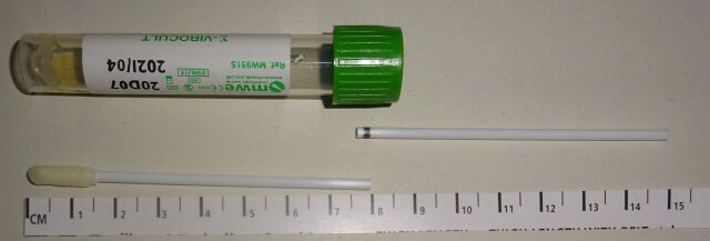
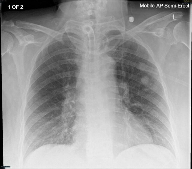
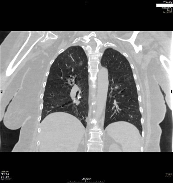
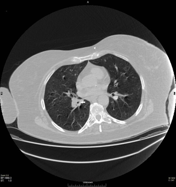
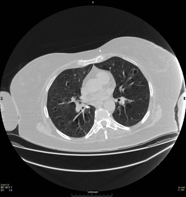
We report the case of a bronchial foreign body, following a tracheostomy site swab for SARS-CoV-2, aiming to raise awareness and vigilance. A qualified nurse was performing a routine SARS-CoV-2 swab on a 51-year-old woman, fitted with a tracheostomy in the recent past following a craniotomy. This was part of the discharging protocol to a nursing home. During the sampling, part of the swab stylet snapped and was inadvertently dropped through the tracheostomy site. Initial CT imaging was reported as showing no signs of a foreign body but some inflammatory changes. Bedside flexible endoscopy through the tracheostomy site revealed the swab in a right lobar bronchus. This was subsequently removed by flexible bronchoscopy. This case highlights the need for clear guidance on how samples for SARS-CoV-2 are taken from patients with front of neck airways (laryngectomy/tracheοstomy) and the potential pitfalls involved.
Keywords: ear; medical-surgical nursing; nose and throat/otolaryngology; otolaryngology / ENT; radiology.
© BMJ Publishing Group Limited 2020. No commercial re-use. See rights and permissions. Published by BMJ.
Conflict of interest statement
Competing interests: None declared.
Figures
References
-
- World Health Organization . Clinical management of severe acute respiratory infection (SARI) when COVID-19 disease is suspected: interim guidance, 13 March 2020 (NO. WHO/2019-nCoV/clinical/2020.4). World Health Organization, 2020.
-
- Centers for Disease Control and Prevention . Interim guidelines for collecting, handling, and testing clinical specimens from persons under investigation (PUIs) for coronavirus disease 2019 (COVID-19)..
-
- NTSP . NTSP considerations for tracheostomy in the COVID-19 outbreak. Available: http://www.tracheostomy.org.uk/storage/files/NTSP%20COVID_19%20tracheost...
Publication types
MeSH terms
LinkOut - more resources
Full Text Sources
Medical
Research Materials
Miscellaneous