Case Reports
. 2020 Aug 27;13(8):e235790.
doi: 10.1136/bcr-2020-235790.

Haemorrhagic encephalitis in the garb of scrub typhus

Affiliations
Case Reports

Haemorrhagic encephalitis in the garb of scrub typhus

Saurabh Gaba et al. BMJ Case Rep. .

Abstract

A 19-year-old girl presented with fever, headache, vomiting and drowsiness. She had grade 1 papilloedema and neck rigidity but no focal deficits or seizures. Cerebrospinal fluid analysis revealed lymphocytic pleocytosis, slightly elevated protein and normal glucose. MRI of the brain showed a hyperintense lesion in left ganglio-capsular region on the fluid attenuation inversion recovery sequence with perilesional oedema and mild midline shift. Haemorrhage was seen in the region on susceptibility weighted imaging . The patient was thoroughly investigated for known causes of meningoencephalitis, but the diagnosis of scrub typhus was delayed till the 10th day of illness. She was treated with doxycycline for 2 weeks and had marked improvement, both clinically and radiologically. Literature review has revealed that although meningoencephalitis in scrub typhus is not uncommon, such atypical lesions on brain MRI are a rarity. Serial imaging was performed to document the disease progression and resolution on treatment.

Keywords: infection (neurology); infectious diseases; tropical medicine (infectious disease).


Conflict of interest statement

Competing interests: None declared.

Figures

Figure 1
Figure 1
MRI on 6th day of illness (before doxycycline)—axial sections of T1 weighted image (A), gradient echo sequence (GRE) (B) and T2 weighted image (C). Large ill-defined area of altered signal intensity diffusely involving the left basal ganglia, thalamus and internal capsule. It is isointense to hypointense on T1 and hyperintense on T2 weighted images. There is mild perilesional oedema and mass effect is seen in the form of partial effacement of left lateral and third ventricles. There is mild dilatation of right lateral ventricle and mild midline shift to right side. The GRE sequence shows punctate foci with minimal blooming in left ganglio-capsular region, suggestive of nominal haemorrhage.
Figure 2
Figure 2
Fundus showing grade 1 papilloedema. The nasal margin (arrow) of the optic discs is blurred and the temporal margin is well defined.
Figure 3
Figure 3
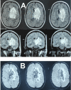
MRI on 9th day of illness (before doxycycline)—axial sections of fluid attenuation inversion recovery (FLAIR) sequence in upper row and coronal sections in the middle row (A), and axial sections of susceptibility weighted imaging (SWI) (B). Altered signal intensity appearing heterogeneously hyperintense on FLAIR sequence is seen in left ganglio-capsular region and thalamus. The lesion has irregular peripheral enhancement and enhancement is also seen in ependymal surface of left lateral ventricle. The lesion has moderate surrounding oedema which also involves genu and body of corpus callosum on left side, left cerebral peduncle and right thalamus. Mass effect is seen in the form of effacement of body and frontal region of left lateral ventricle and third ventricle. There is mild midline shift. SWI sequence shows extensive area of blooming in the left basal ganglia region, suggestive of significant haemorrhage.
Figure 4
Figure 4
MRI on 17th day of illness (7th day of treatment with doxycycline)—axial sections of susceptibility weighted imaging sequence showing markedly reduced blooming and oedema in the left ganglio-capsular region. There is no midline shift.

Similar articles

Cited by

References

    1. Kastrup O, Wanke I, Maschke M. Neuroimaging of infections. NeuroRx 2005;2:324–32. 10.1602/neurorx.2.2.324 - DOI - PMC - PubMed
    1. Jayaraman K, Rangasami R, Chandrasekharan A. Magnetic resonance imaging findings in viral encephalitis: a pictorial essay. J Neurosci Rural Pract 2018;9:556–60. 10.4103/jnrp.jnrp_120_18 - DOI - PMC - PubMed
    1. Thurnher MM, Thurnher SA, Schindler E. CNS involvement in AIDS: spectrum of CT and MR findings. Eur Radiol 1997;7:1091–7. 10.1007/s003300050260 - DOI - PubMed
    1. Duarte SBL, Oshima MM, Mesquita JVdoA, et al. . Magnetic resonance imaging findings in central nervous system cryptococcosis: comparison between immunocompetent and immunocompromised patients. Radiol Bras 2017;50:359–65. 10.1590/0100-3984.2016.0017 - DOI - PMC - PubMed
    1. Jayakrishnan MP, Krishnakumar P. Clinical profile of acute disseminated encephalomyelitis in children. J Pediatr Neurosci 2010;5:111–4. 10.4103/1817-1745.76098 - DOI - PMC - PubMed

Publication types