. 2020 Aug 28;10(1):14236.
doi: 10.1038/s41598-020-71127-2.

Comparison of 68Ga-DOTANOC with 18F-FDG using PET/MRI imaging in patients with pulmonary tuberculosis

Affiliations

Comparison of 68Ga-DOTANOC with 18F-FDG using PET/MRI imaging in patients with pulmonary tuberculosis

Claire M Naftalin et al. Sci Rep. .

Abstract

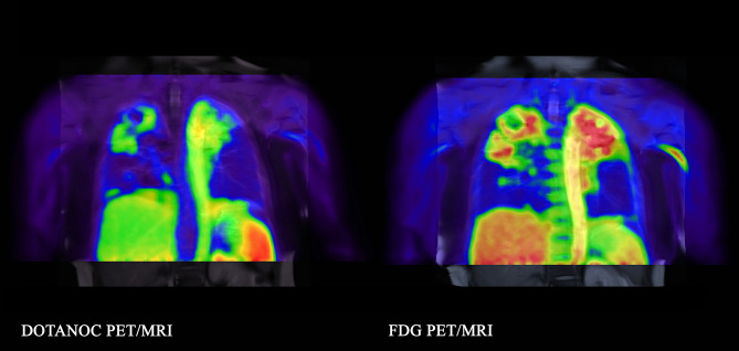
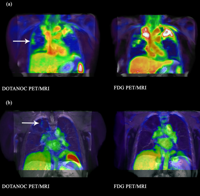
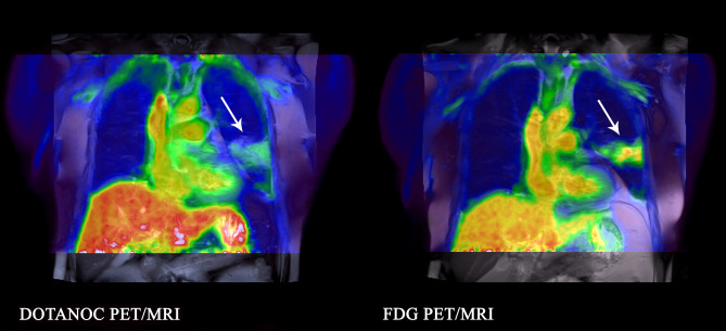
We compared the somatostatin analog radioligand, DOTANOC, with FDG, to determine whether there was increased detection of active or sub-clinical lesions in pulmonary tuberculosis (TB) with DOTANOC. Three groups were recruited: (1) active pulmonary TB; (2) IGRA-positive household TB contacts; (3) pneumonia (non-TB). DOTANOC PET/MRI followed by FDG PET/MRI was performed in active TB and pneumonia groups. TB contacts underwent FDG PET/MRI, then DOTANOC PET/MRI if abnormalities were detected. Quantitative and qualitative analyses were performed for total lung and individual lesions. Eight active TB participants, three TB contacts and three pneumonia patients had paired PET/MRI scans. In the active TB group, median SUVmax[FDG] for parenchymal lesions was 7.69 (range 3.00-15.88); median SUVmax[DOTANOC] was 2.59 (1.48-6.40). Regions of tracer uptake were fairly similar for both radioligands, albeit more diffusely distributed in the FDG scans. In TB contacts, two PET/MRIs had parenchymal lesions detected with FDG (SUVmax 5.50 and 1.82), with corresponding DOTANOC uptake < 1. FDG and DOTANOC uptake was similar in pneumonia patients (SUVmax[FDG] 4.17-6.18; SUVmax[DOTANOC] 2.92-4.78). DOTANOC can detect pulmonary TB lesions, but FDG is more sensitive for both active and sub-clinical lesions. FDG remains the preferred ligand for clinical studies, although DOTANOC may provide additional value for pathogenesis studies.


Conflict of interest statement

The authors declare no competing interests.

Figures

Figure 1
Figure 1
Example of diffuse uptake of FDG (SUVmax 8.22) compared to DOTANOC uptake (SUVmax 2.96) which is more discrete. 55-year-old woman with pulmonary TB, smear 3+, on Day 8 of TB treatment.
Figure 2
Figure 2
Lesions visualised with FDG but not DOTANOC in pulmonary TB (a) Lesions in right upper lobe visualised with FDG, largest volume 16.5 cm3, SUVmax 2.89. No uptake with DOTANOC. 22-year-old male, smear 2+, on TB treatment for 28 days. (b) Lesion in posterior basal segment of right lower lobe visualised with FDG, volume 4.3 cm3, SUVmax 2.50. No uptake with DOTANOC. 29-year-old male, smear 3+, on TB treatment for 25 days.
Figure 3
Figure 3
Lesions visualised with DOTANOC but not FDG in pulmonary TB (a) Lesion in right middle lobe visualised with DOTANOC, volume 0.6 cm3, SUVmax 1.92. No uptake with FDG. 55-year-old woman, smear 3+, on Day 8 of TB treatment. (b) Lesion at apex right upper lobe visualised with DOTANOC, volume 0.4 cm3, SUVmax 1.80. No uptake with FDG. 57-year-old man, smear negative, on Day 11 of TB treatment.
Figure 4
Figure 4
Lesion visualised in anterior segment of the left upper lobe adjacent to the aorta with FDG in 47-year-old female, IGRA-positive TB contact, taking isoniazid for 5 days. FDG SUVmax 5.50. No DOTANOC uptake.
Figure 5
Figure 5
Similar distribution of tracer uptake of DOTANOC and FDG in a 73-year old male patient with non-TB pneumonia. Lesion visualised in the left lower lobe mainly involving the superior segment.

References

    1. World Health Organisation. Global tuberculosis report 2019. https://apps.who.int/iris/bitstream/handle/10665/329368/9789241565714-en... (2019).
    1. Thomas BA, et al. A comparison of 18F-FDG PET/MR with PET/CT in pulmonary tuberculosis. Nucl. Med. Commun. 2017;38:971–978. doi: 10.1097/mnm.0000000000000743. - DOI - PubMed
    1. Chen RY, et al. PET/CT imaging correlates with treatment outcome in patients with multidrug-resistant tuberculosis. Sci. Transl. Med. 2014;6:265ra166. doi: 10.1126/scitranslmed.3009501. - DOI - PMC - PubMed
    1. Esmail H, et al. Characterization of progressive HIV-associated tuberculosis using 2-deoxy-2-[18F]fluoro-d-glucose positron emission and computed tomography. Nat. Med. 2016;22:1090–1093. doi: 10.1038/nm.4161. - DOI - PMC - PubMed
    1. Malherbe ST, et al. Persisting positron emission tomography lesion activity and mycobacterium tuberculosis mRNA after tuberculosis cure. Nat. Med. 2016;22:1094–1100. doi: 10.1038/nm.4177. - DOI - PMC - PubMed

Publication types

MeSH terms