. 2020 Dec;237(12):3653-3664.
doi: 10.1007/s00213-020-05645-2. Epub 2020 Aug 28.

Dorsal hippocampal interleukin-1 signaling mediates heroin withdrawal-enhanced fear learning

Affiliations

Dorsal hippocampal interleukin-1 signaling mediates heroin withdrawal-enhanced fear learning

Shveta V Parekh et al. Psychopharmacology (Berl). 2020 Dec.

Abstract

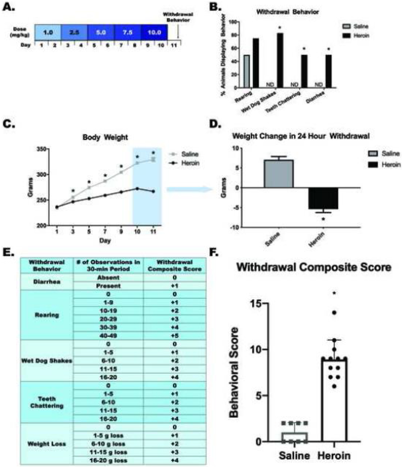
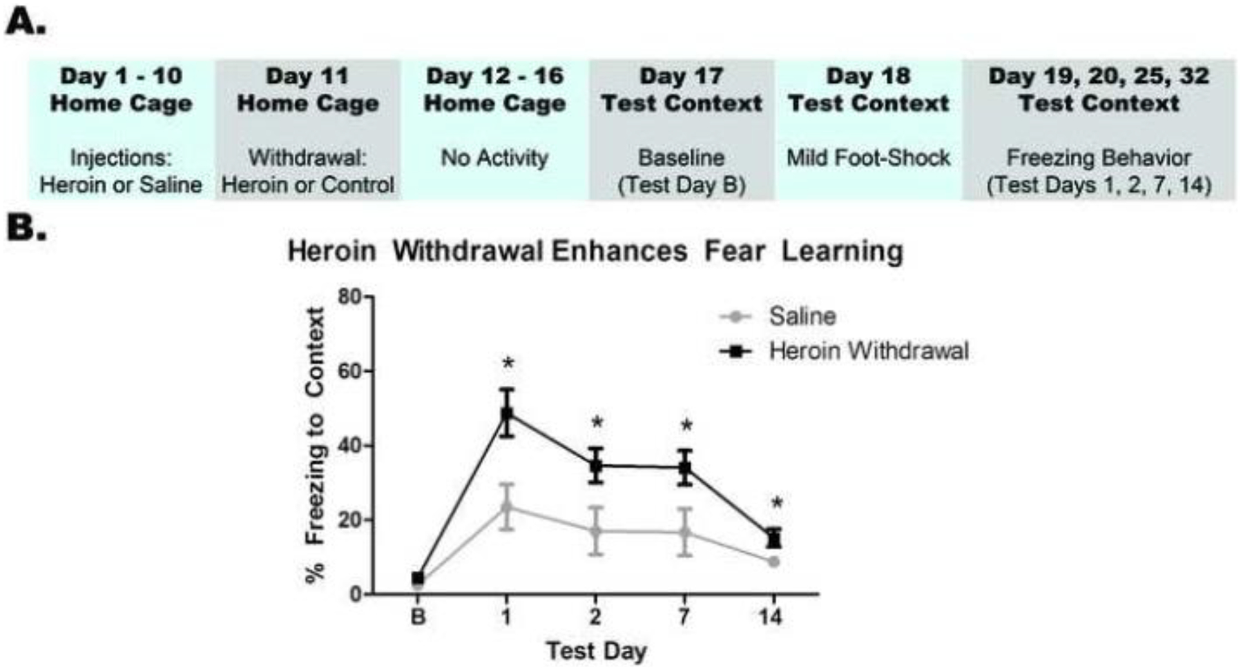
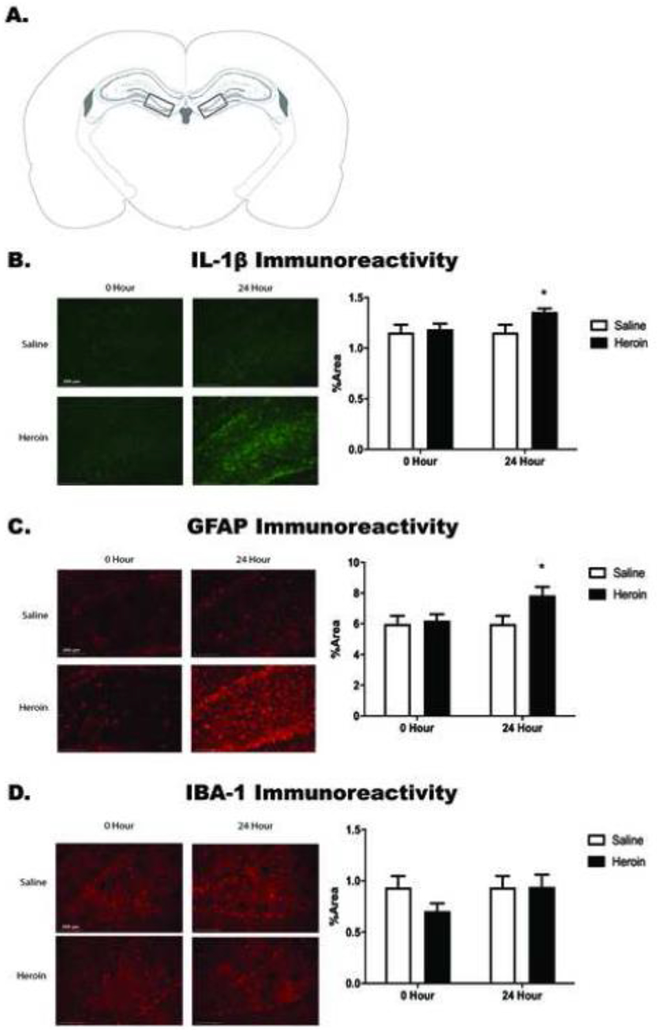
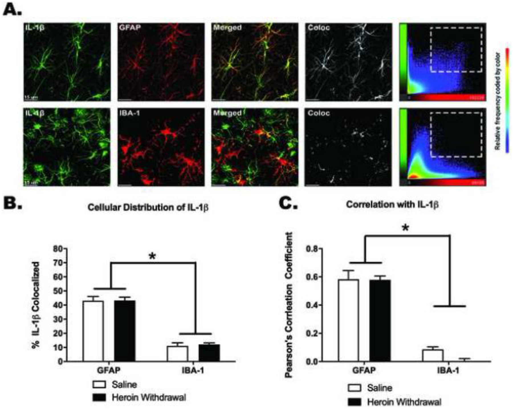
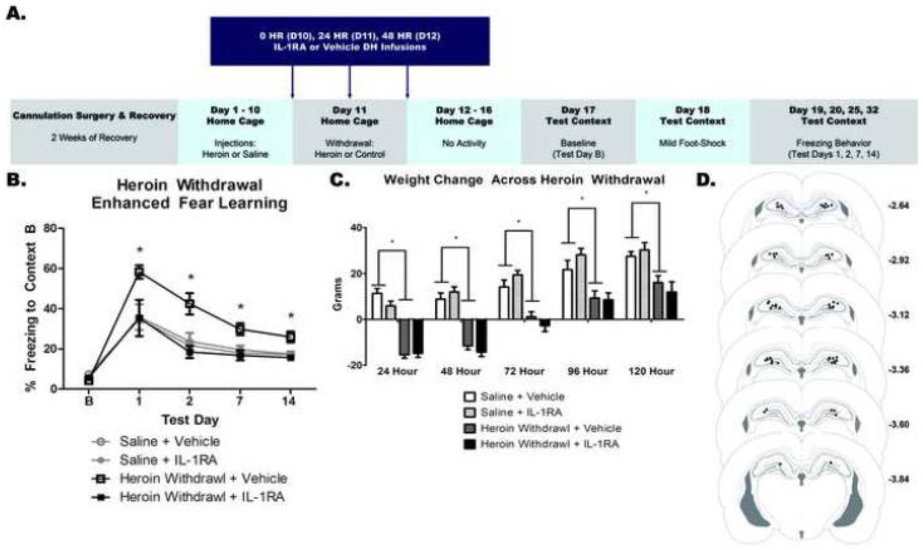
Converging evidence suggests opioid abuse can increase the incidence and severity of post-traumatic stress disorder (PTSD) in clinical populations. Interestingly, opioid withdrawal alone can produce symptoms similar to those of PTSD. Despite this association, the neural mechanisms underlying the relationship of opioid abuse, withdrawal, and PTSD is poorly understood. Our laboratory has investigated the neurobiological underpinnings of stress-enhanced fear learning (SEFL), an animal model of PTSD-like symptoms. We have previously shown that, in SEFL, a severe footshock induces an increase in dorsal hippocampal (DH) interleukin-1β (IL-1β), and subsequent fear learning is blocked by DH IL-1 receptor antagonism (IL-1RA). Given that opioids and stress engage similar neuroimmune mechanisms, the present experiments investigate whether the same mechanisms drive heroin withdrawal to induce a PTSD-like phenotype. First, we tested the effect of a chronic escalating heroin dose and withdrawal regimen on fear learning and found it produces enhanced future fear learning. Heroin withdrawal also induces a time-dependent, region-specific increase in IL-1β and glial fibrillary acidic protein (GFAP) immunoreactivity within the dentate gyrus of the DH. IL-1β was significantly colocalized with GFAP, indicating astrocytes may be involved in increased IL-1β. Moreover, intra-DH infusions of IL-1RA 0, 24, and 48 h into heroin withdrawal prevents the development of enhanced fear learning but does not alter withdrawal-induced weight loss. Collectively, our data suggests heroin withdrawal is sufficient to produce enhanced fear learning, astrocytes may play a role in heroin withdrawal-induced IL-1β, and DH IL-1 signaling during withdrawal mediates the development of heroin withdrawal-enhanced fear learning.

Keywords: Glia; Heroin; Neuroimmunology; Opioid; PTSD; Withdrawal.


Conflict of interest statement

Conflict of Interest Statement: On behalf of all authors, the corresponding author states that there is no conflict of interest.

Figures

Figure 1.
Figure 1.. Chronic escalating heroin administration produces withdrawal behaviors.
Chronic escalating heroin administration schematic (A). Withdrawal behavior (B). Overall body weight across days of heroin administration (C) and weight change in 24-hour withdrawal (D) Withdrawal composite score explanation chart (E). Heroin animals have a higher withdrawal composite score (range between 6 and 14) than saline animals (range between 0 and 2) (F). *, statistically significant difference relative to respective control. Error bars indicate SEM.
Figure 2.
Figure 2.. Heroin withdrawal causes enhanced fear learning.
Experimental timeline (A). Heroin withdrawal significantly enhances contextual freezing behavior to a single mild footshock (B). *, statistically significant difference relative to respective control. Error bars indicate SEM.
Figure 3.
Figure 3.. Heroin withdrawal increases Il-1β and GFAP immunoreactivity in the dentate gyrus of the dorsal hippocampus.
Paxinos and Watson (2007) schematic depicting bilateral image acquisition location. Each rectangular box represents the area in which images were taken within the DG of DH (A). Representative images (20X) for saline and heroin animals at both 0- and 24-hour time points (left) and quantification of positive fluorescence stain (right) of IL-1β (B), GFAP (C), and IBA-1 (D) taken within the DG of DH. *, statistically significant difference relative to respective control. Error bars indicate SEM.
Figure 4.
Figure 4.. IL-1β signal is colocalized with GFAP, and not with IBA-1, in the dentate gyrus of the dorsal hippocampus in heroin withdrawn and control animals.
Representative 63X images of the DG. Images were taken in the area as seen in Figure 3A. Colocalization scatter plots show the signal intensity for each voxel in the Z-stack. (A). Colocalization of IL-1β signal with GFAP (top row) and IBA-1 (bottom row), respectively (B). Pearson’s correlation coefficient of colocalization of IL-1β signal intensity with GFAP and IBA-1 signal intensity, respectively (C). *, statistically significant difference relative to respective control. Error bars indicate SEM. Background signal was subtracted using Bitplane Imaris.
Figure 5.
Figure 5.. IL-1RA prevents the development of heroin withdrawal-enhanced fear learning but not weight loss
Experimental timeline (A). DH IL-1RA significantly attenuated HW-EFL (B). DH IL-1RA had no effect on weight change (C). Paxinos and Watson (2007) schematic with DH cannula placements shown. Each circle represents termination site of the cannula tract (D). *, statistically significant difference relative to respective control. Error bars indicate SEM.

References

    1. Barres BA (2008) The mystery and magic of glia: a perspective on their roles in health and disease Neuron 60:430–440 doi:10.1016/j.neuron.2008.10.013 - DOI - PubMed
    1. Benediktsson AM, Schachtele SJ, Green SH, Dailey ME (2005) Ballistic labeling and dynamic imaging of astrocytes in organotypic hippocampal slice cultures J Neurosci Methods 141:41–53 doi:10.1016/j.jneumeth.2004.05.013 - DOI - PubMed
    1. Bennett AS, Elliott L, Golub A, Wolfson-Stofko B, Guarino H (2017) Opioid-Involved Overdose Among Male Afghanistan/Iraq-Era U.S. Military Veterans: A Multidimensional Perspective Subst Use Misuse 52:1701–1711 doi:10.1080/10826084.2017.1306563 - DOI - PMC - PubMed
    1. Bland ST, Hutchinson MR, Maier SF, Watkins LR, Johnson KW (2009) The glial activation inhibitor AV411 reduces morphine-induced nucleus accumbens dopamine release Brain Behav Immun 23:492–497 doi:10.1016/j.bbi.2009.01.014 - DOI - PMC - PubMed
    1. Block ML, Zecca L, Hong JS (2007) Microglia-mediated neurotoxicity: uncovering the molecular mechanisms Nat Rev Neurosci 8:57–69 doi:10.1038/nrn2038 - DOI - PubMed

MeSH terms

LinkOut - more resources