. 2020 Dec 15;88(1):202-210.
doi: 10.1093/neuros/nyaa355.

A Prognostic Gene-Expression Signature and Risk Score for Meningioma Recurrence After Resection

Affiliations

A Prognostic Gene-Expression Signature and Risk Score for Meningioma Recurrence After Resection

William C Chen et al. Neurosurgery. .

Abstract

Background: Prognostic markers for meningioma are needed to risk-stratify patients and guide postoperative surveillance and adjuvant therapy.

Objective: To identify a prognostic gene signature for meningioma recurrence and mortality after resection using targeted gene-expression analysis.

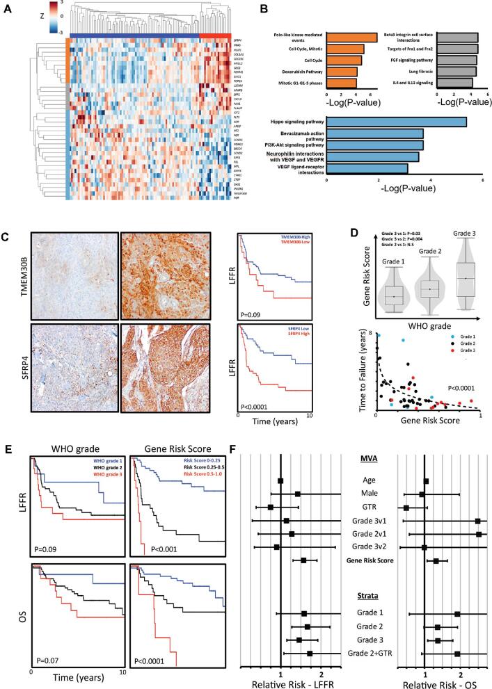
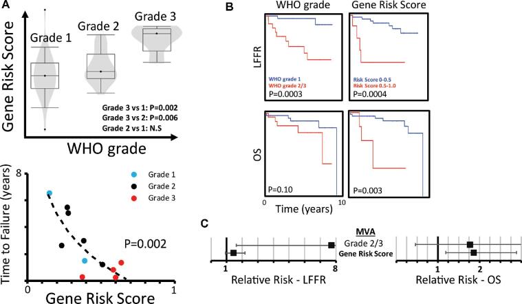
Methods: Targeted gene-expression analysis was used to interrogate a discovery cohort of 96 meningiomas and an independent validation cohort of 56 meningiomas with comprehensive clinical follow-up data from separate institutions. Bioinformatic analysis was used to identify prognostic genes and generate a gene-signature risk score between 0 and 1 for local recurrence.

Results: We identified a 36-gene signature of meningioma recurrence after resection that achieved an area under the curve of 0.86 in identifying tumors at risk for adverse clinical outcomes. The gene-signature risk score compared favorably to World Health Organization (WHO) grade in stratifying cases by local freedom from recurrence (LFFR, P < .001 vs .09, log-rank test), shorter time to failure (TTF, F-test, P < .0001), and overall survival (OS, P < .0001 vs .07) and was independently associated with worse LFFR (relative risk [RR] 1.56, 95% CI 1.30-1.90) and OS (RR 1.32, 95% CI 1.07-1.64), after adjusting for clinical covariates. When tested on an independent validation cohort, the gene-signature risk score remained associated with shorter TTF (F-test, P = .002), compared favorably to WHO grade in stratifying cases by OS (P = .003 vs P = .10), and was significantly associated with worse OS (RR 1.86, 95% CI 1.19-2.88) on multivariate analysis.

Conclusion: The prognostic meningioma gene-expression signature and risk score presented may be useful for identifying patients at risk for recurrence.

Keywords: Biomarker; Expression; Gene; Gene expression; Meningioma; Prognostic; Radiation; Recurrence; Resection; Survival; WHO grade.


Figures

FIGURE 1.
FIGURE 1.
Targeted gene-expression analysis of clinically aggressive meningiomas identifies a prognostic gene signature. A, Unsupervised hierarchical clustering of prognostic genes identified using PAM confirms the ability of the gene set to stratify meningioma patients into high-risk (red cluster) and lower-risk categories (blue cluster, log-rank test, P < .0001). Gene expression is normalized by row. B, Gene enrichment analysis of prognostic gene clusters from A identifies a tightly correlated set of genes involved in cell-cycle processes (orange cluster), and clusters of genes involved in cellular signaling and extracellular matrix interactions (light blue and grey clusters). C, Representative IHC images demonstrating high TMEM30B staining on the top right (20x magnification) and low/absent TMEM30B staining on the top left. Similarly, representative IHC images demonstrating low SFRP4 staining (20x magnification) on the bottom left and high SFRP4 staining on the bottom right are shown. Low TMEM30B staining (15 of 96 meningiomas, 16%) is associated with a trend towards worse LFFR, and high SFRP4 staining (46 of 94 meningiomas, 49%) is significantly associated with worse LFFR. D, Elastic net regression was used to generate a gene-signature risk score between 0 and 1 per tumor sample (accuracy 0.80, AUC 0.86). Gene risk score correlates with tumor grade and is correlated with a faster time to failure (TTF) (TTF vs log(gene risk), P < .0001, F-test). Meningiomas with a gene risk score of greater than 0.5 uniformly recur within 2 yr of resection. Meningiomas which did not recur are not plotted. E, The gene-signature risk score outperforms WHO grade in stratifying LFFR (P < .001 vs P = .09, log-rank test) and OS (P < .0001 vs P = .07, log-rank test). F, After adjusting for age, sex, extent of resection, and grade using multivariate Cox regression, the gene-signature risk score is independently associated with recurrence (RR 1.56 per 0.1 risk score increase, 95% CI 1.30-1.90) and mortality (RR 1.32 per 0.1 increase, 95% CI 1.07-1.64). After stratifying patients by grade, the gene-signature risk score remains significantly prognostic for meningioma recurrence and mortality on univariate Cox regression. Further, among gross totally resected grade 2 tumors (Grade 2 + gross total resection (GTR)), the gene risk score is significantly prognostic of recurrence.
FIGURE 2.
FIGURE 2.
Prognostic gene-signature risk score validation in an independent dataset. A, Meningioma gene-signature risk scores were calculated on an independent validation dataset from an outside institution. The gene-signature risk score remains correlated with WHO grade and with faster TTF among patients who recurred (TTF vs log(GS risk), P = .002, F-test). Meningiomas which did not recur are not plotted. B, The gene risk score remains significantly associated with worse LFFR (P = .0004, log-rank test) and compares favorably to WHO grade in stratifying patients by OS (P = .003 vs P = .10, log-rank test). C, The gene-signature risk score remains significantly prognostic for mortality (RR 1.86 per 0.1 increase, 95% CI 1.19-2.88) after adjusting for WHO grade on Cox regression.

References

    1. Ostrom QT, Gittleman H, Truitt G, Boscia A, Kruchko C, Barnholtz-Sloan JS. CBTRUS statistical report: primary brain and other central nervous system tumors diagnosed in the united states in 2011-2015. Neuro Oncol. 2018;20(Suppl_4):iv1-iv86. - PMC - PubMed
    1. Aizer AA, Bi WL, Kandola MS et al. . Extent of resection and overall survival for patients with atypical and malignant meningioma. Cancer. 2015;121(24):4376-4378. - PubMed
    1. Chen WC, Magill ST, Wu A et al. . Histopathologic features predictive of local control of atypical meningioma after surgery and adjuvant radiotherapy. J Neurosurg. 2018;130(2):443-450. - PubMed
    1. Condra KS, Buatti JM, Mendenhall WM, Friedman WA, Marcus RB, Rhoton AL. Benign meningiomas: primary treatment selection affects survival. Int J Radiat Oncol Biol Phys. 1997;39(2):427-36. - PubMed
    1. Cahill KS, Claus EB.. Treatment and survival of patients with nonmalignant intracranial meningioma: results from the surveillance, epidemiology, and end results program of the national cancer institute. Clinical article. J Neurosurg. 2011;115(2):259-67. - PMC - PubMed

Publication types