. 2020 Sep 18;432(20):5665-5680.
doi: 10.1016/j.jmb.2020.08.017. Epub 2020 Aug 26.

The Multidrug Transporter MdfA Deviates from the Canonical Model of Alternating Access of MFS Transporters

Affiliations

The Multidrug Transporter MdfA Deviates from the Canonical Model of Alternating Access of MFS Transporters

Eliane H Yardeni et al. J Mol Biol. .

Abstract

The prototypic multidrug (Mdr) transporter MdfA from Escherichia coli efflux chemically- dissimilar substrates in exchange for protons. Similar to other transporters, MdfA purportedly functions by alternating access of a central substrate binding pocket to either side of the membrane. Accordingly, MdfA should open at the cytoplasmic side and/or laterally toward the membrane to enable access of drugs into its pocket. At the end of the cycle, the periplasmic side is expected to open to release drugs. Two distinct conformations of MdfA have been captured by X-ray crystallography: An outward open (Oo) conformation, stabilized by a Fab fragment, and a ligand-bound inward-facing (If) conformation, possibly stabilized by a mutation (Q131R). Here, we investigated how these structures relate to ligand-dependent conformational dynamics of MdfA in lipid bilayers. For this purpose, we combined distances measured by double electron-electron resonance (DEER) between pairs of spin labels in MdfA, reconstituted in nanodiscs, with cysteine cross-linking of natively expressed membrane-embedded MdfA variants. Our results suggest that in a membrane environment, MdfA assumes a relatively flexible, outward-closed/inward-closed (Oc/Ic) conformation. Unexpectedly, our data show that neither the substrate TPP nor protonation induces large-scale conformational changes. Rather, we identified a substrate-responsive lateral gate, which is open toward the inner leaflet of the membrane but closes upon drug binding. Together, our results suggest a modified model for the functional conformational cycle of MdfA that does not invoke canonical elements of alternating access.

Keywords: DEER; MdfA; conformational cycle; multidrug transport; nanodiscs.


Figures

Figure 1.
Figure 1.. Distance distributions for pairs on the cytoplasmic side of MdfA.
A cytoplasmic view of MdfA in the (a) If (pdb 4ZP0) and (b) OO (pdb 6GV1) crystal structures.Residues that were labeled with MTSSL are marked as red spheres and pairs of residues are connected by red lines. (c - i). Distance distributions of the apo protein were measured at pH 7.5 (blue) and 9.0 (orange). The MMM-derived distance distributions, calculated using the crystal structures 4ZP0 (If, shaded purple) and 6GV1 (OO, shaded green), overlaid with the DEER derived distance distributions at pH 7.5 (blue) and pH 9.0 (orange) [12]. Mutant pairs are indicated above their corresponding graphs. Mutant TM120-TM8280 (i), was solubilized with α-DDM. Asterisks indicate peaks resulting from partial aggregation observed in some samples.
Figure 2.
Figure 2.. Distance distributions for pairs on the periplasmic side of MdfA.
A periplasmic view of MdfA in the (a) If (pdb 4ZP0) and (b) OO (pdb 6GV1) crystal structures. Residues that were labeled with MTSSL are marked as red spheres and pairs of residues are connected by red lines. (c - k). Distance distributions of the apo protein were measured at pH 7.5 (blue) and 9.0 (orange). The MMM-derived distance distributions, calculated using the crystal structures 4ZP0 (If, shaded purple) and 6GV1 (OO, shaded green), overlaid with the DEER derived distance distributions at pH 7.5 (blue) and pH 9.0 (orange) [12]. Mutant pairs are indicated above their corresponding graphs. Asterisks indicate peaks resulting from partial aggregation observed in some samples.
Figure 3.
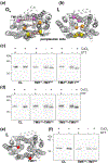
Figure 3.. Disulfide bond formation on the periplasmic side of a cell membrane-embedded MdfA.
(a - b). The periplasmic face of MdfA in the (a) OO (pdb 6GV1) and (b) If (pdb 4ZP0) crystal structures. Cysteine insertions are marked in pink (TM255-TM11369), orange (TM141-TM7243) and yellow (TM5162-TM8254/TM8258) and the pairs are connected by red lines. (c - d). Membranes from cells expressing CL-MdfA (CL), (c) TM255-TM11369 and TM141-TM7243, (d) TM5162-TM8254 and TM5162- TM8258, were incubated with or without CuCl2, separated by a 15% SDS-PAGE and analyzed by Western blot. (e) The periplasmic face of MdfA in the If (pdb 4ZP0) conformation. Cysteine insertions are marked as red spheres. (f) Membranes from cells expressing CL-MdfA, or TM5163-TM7245 were incubated with or without CuCl2, separated by a 10% SDS-PAGE and analyzed by Western blot.
Figure 4.
Figure 4.. Protonation-induced distance changes of MdfA.
Distance distributions of the apo protein at pH 7.5 (blue) and 5.0 (brown) of mutants in the (a) periplasmic side and (c) cytoplasmic side of MdfA [12]. Mutant TM5140-TM11348 was solubilized with α-DDM. Asterisks indicate peaks resulting from partial aggregation observed in some samples. (b) The periplasmic face of the If crystal structure of MdfA (pdb 4ZP0) is shown with TM1 and TM7 (orange segments) and residues 46 and 246 (blue spheres).
Figure 5.
Figure 5.. TPP-induced distance changes of MdfA.
Distance distributions measured at pH 7.5 of the apo protein (blue) and in the presence of TPP (purple) of (a) periplasmically- and (b) cytoplasmically-labeled MdfA [12]. Mutant TM5140-TM11348 was solubilized with α- DDM. Asterisks indicate peaks resulting from partial aggregation observed in some samples.
Figure 6.
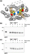
Figure 6.. Disulfide bond formation between TM1 and TM7 in a membrane-embedded MdfA.
(a) The periplasmic face of MdfA in the If (pdb 4ZP0) crystal structures. TM1 and TM7 are shown in yellow and orange. The double-cysteine pairs TM141-TM7243 and TM146- TM7246 are shown as blue and red spheres, respectively. (b - c) Membranes from cells expressing (b) TM141-TM7243 and (c) TM146-TM7246, were incubated with or without TPP, followed by an additional incubation at 4°C, for 0–8 min, in the presence of CuCl2. Samples were then separated on an SDS-PAGE and analyzed by Western blot.
Figure 7.
Figure 7.. Disulfide bond formation between cysteines inserted on both sides of a putative inner leaflet lateral gate(s) of MdfA.
The If (4ZP0, left panels) and the OO (6GV1, right panels) crystal structures of MdfA with (a) TM5 and TM8 shown in blue and cysteine insertions marked as red spheres and (e) TM2 and TM11 shown in green and cysteine insertions marked as yellow spheres. Membranes from cells expressing (b) CL- MdfA (CL), TM5140-TM8276, (c) CL-MdfA (CL), TM5144-TM8272, (d) TM5144-TM8272, or (f) TM273-TM11347 were incubated with or without CuCl2, separated by (b) 15%, (c - d, f) 12.5% SDS-PAGE and analyzed by Western blotting.
Figure 8.
Figure 8.. Model for MdfA-catalyzed substrate/H+ antiport.
1. In the proposed resting state of membrane-embedded MdfA, the protein is deprotonated. Both the periplasmic and cytoplasmic sides are relatively closed. 2. A lipophilic drug (LD) may enter the multidrug recognition pocket from the inner leaflet of the membrane, between TM5 and TM8, while less hydrophobic drugs (hydrophilic drugs, HD) may enter from the cytoplasm, between TM4 and TM10. 3. Substrate binding triggers a subtle conformational change that further closes the cytoplasmic side and the periplasmic edges of TM1 and TM7, while slightly exposing the multidrug-binding pocket to the periplasm. 4. Protons bind to MdfA once the substrate is released to the extracellular side. 5. The proton-binding event triggers a conformational switch and proton-release recycles MdfA to the resting state.

References

    1. Saier MH Jr., Eng BH, Fard S, Garg J, Haggerty DA, Hutchinson WJ, et al. Phylogenetic characterization of novel transport protein families revealed by genome analyses. Biochim Biophys Acta. 1999;1422:1–56. - PubMed
    1. Fluman N, Bibi E. Bacterial multidrug transport through the lens of the major facilitator superfamily. Biochim Biophys Acta. 2009;1794:738–47. - PubMed
    1. Yardeni EH, Zomot E, Bibi E. The fascinating but mysterious mechanistic aspects of multidrug transport by MdfA from Escherichia coli. Res Microbiol. 2017. - PubMed
    1. Adler J, Bibi E. Membrane topology of the multidrug transporter MdfA: complementary gene fusion studies reveal a nonessential C-terminal domain. J Bacteriol. 2002;184:3313–20. - PMC - PubMed
    1. Sigal N, Vardy E, Molshanski-Mor S, Eitan A, Pilpel Y, Schuldiner S, et al. 3D model of the Escherichia coli multidrug transporter MdfA reveals an essential membrane-embedded positive charge. Biochemistry. 2005;44:14870–80. - PubMed

Publication types

LinkOut - more resources