. 2021 Apr 23;223(8):1488-1496.
doi: 10.1093/infdis/jiaa548.

Hemolytic Membrane Vesicles of Group B Streptococcus Promote Infection

Affiliations

Hemolytic Membrane Vesicles of Group B Streptococcus Promote Infection

Blair Armistead et al. J Infect Dis. .

Abstract

Background: Group B streptococci (GBS) are β-hemolytic, Gram-positive bacteria associated with fetal injury, preterm birth, spontaneous abortion, and neonatal infections. A key factor promoting GBS virulence is the β-hemolysin/cytolysin, a pigmented ornithine rhamnolipid (also known as granadaene) associated with the bacterial surface.

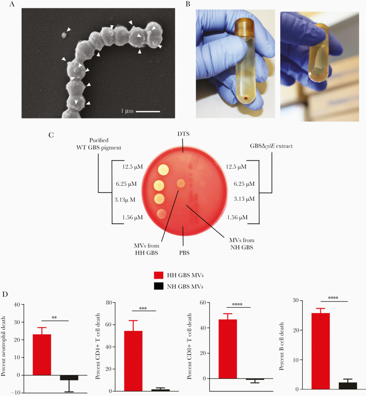
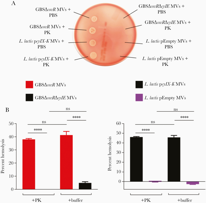
Methods: A previous study indicated that GBS produce small structures known as membrane vesicles (MVs), which contain virulence-associated proteins. In this study, we show that GBS MVs are pigmented and hemolytic, indicating that granadaene is functionally active in MVs.

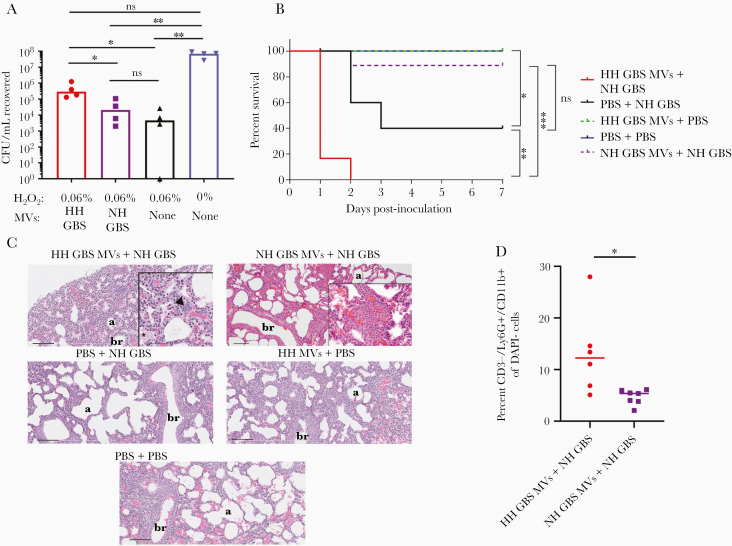
Results: In addition, MVs from hyperhemolytic GBS induced greater cell death of neutrophils, T cells, and B cells compared with MVs from isogenic nonhemolytic GBS, implicating MVs as a potential mechanism for granadaene-mediated virulence. Finally, hemolytic MVs reduced oxidative killing of GBS and aggravated morbidity and mortality of neonatal mice infected with GBS.

Conclusions: These studies, taken together, reveal a novel mechanism by which GBS deploy a crucial virulence factor to promote bacterial dissemination and pathogenesis.

Keywords: granadaene; group B streptococcus; hemolysin; immune evasion; membrane vesicles.


Figures

Figure 1.
Figure 1.
Membrane vesicles (MVs) isolated from hemolytic group B streptococci (GBS) are hemolytic and cytolytic. (A) Hyperhemolytic (HH) GBS (GBSΔcovR) were centrifuged, fixed, and analyzed by scanning electron microscopy. Arrowheads indicate MVs, which are seen as spherical structures emerging from the surface of bacterial cells. (B) Pelleted MVs from HH GBS (GBSΔcovR) or nonhemolytic (NH) GBS (GBSΔcovRΔcylE) are shown. (C) Membrane vesicles from HH GBS (GBSΔcovR) or NH GBS (GBSΔcovRΔcylE) were resuspended in phosphate-buffered saline (PBS), sonicated, and 10 µL was spotted onto red blood agar. Purified GBS pigment and equivalent amount of GBSΔcylE extract in DTS at various dilutions were spotted (10 µL) for comparison. (D) Primary human neutrophils, CD4+ T cells, CD8+ T cells, or B cells were incubated with MVs from HH GBS or NH GBS (final concentration = 0.5 mg/mL), and cell death was measured by LDH release into the supernatant relative to Triton X-100 (0.1%)- and PBS-treated controls. Mean and standard error of the mean are shown from 3 experiments performed in technical triplicate. Groups were compared with unpaired, 2-tailed Student’s t test. Neutrophils, P = .0042; CD4+ T cells, P = .0005; CD8+ T cells, P < .0001; B cells, P < .0001. **, P < .01; ***, P < .001; ****, P < .0001.
Figure 2.
Figure 2.
Membrane vesicles (MVs) are hemolytic after treatment with protease. (A) The MVs from GBSΔcovR, GBSΔcovRΔcylE, Lactococcus lactis pcylX-K, or L lactis pEmpty were treated with proteinase K ([PK] final concentration 0.25 mg/mL) or an equivalent volume of phosphate-buffered saline (PBS) for 1 hour at 37°C, and 10 µL of each was spotted on blood agar. (B) The MVs from GBSΔcovR, GBSΔcovRΔcylE, L lactis pcylX-K, or L lactis pEmpty were mixed with human erythrocytes in the presence of PBS or PK (0.25 mg/mL) for 1 hour, and percentage hemolysis was determined relative to Triton X-100 (0.1%)-treated positive controls. One-way analysis of variance with Tukey’s posttest was used. Mean and standard error of the mean from 3 experiments performed in triplicate are shown. ****, P < .0001; ns, P ≥ .05.
Figure 3.
Figure 3.
Hemolytic membrane vesicles (MVs) dampen oxidative killing and exacerbate group B streptococci (GBS) pathogenesis in neonatal mice. (A) Membrane vesicles isolated from hyperhemolytic (HH) GBSΔcovR or nonhemolytic (NH) GBSΔcovRΔcylE were preincubated with 0.06% H2O2 for 45 minutes and then incubated with NH GBS at 37°C for 1 hour. After incubation, remaining H2O2 was quenched with 1000 units of catalase, and surviving colony-forming units (CFU) were enumerated by dilution plating onto TSA. A group consisting of no MVs + H2O2 and no MVs + no H2O2 were included as positive and negative controls, respectively. Treatment groups were compared using a Kruskal-Wallis test with multiple comparisons corrected for false discovery. Hyperhemolytic GBS MVs + H2O2 vs no MVs + H2O2, P = .0170; HH GBS MVs + H2O2 vs NH GBS MVs + H2O2, P = .0395. Nonhemolytic GBS MVs + H2O2 vs no MVs + H2O2, P > .9999; HH GBS MVs + H2O2 vs phosphate-buffered saline (PBS) only, P = .0822; NH GBS MVs + H2O2 vs PBS only, P = .0032; no MVs + H2O2 + PBS, P = .0015. Line at median. (B) Kaplain-Meier plot depicts survival of neonatal mice inoculated with MVs alone or GBS strains alone or GBS strains with MVs, as indicated. Survival between groups was compared using the log-rank test. PBS + PBS (blue) vs PBS + NH GBS (black), P = .0316; PBS + NH GBS (black) vs HH GBS MVs + NH GBS (red), P = .0033; NH GBS MVs + NH GBS (purple) vs HH GBS MVs + NH GBS MVs (red), P = .0002; NH GBS MVs + NH GBS (purple) vs PBS + NH GBS MVs (black), P = .0610. (C) Lungs from neonatal mice in each treatment group were harvested at 24 hours posttreatment, fixed, sectioned, and stained for hematoxylin and eosin. In the HH GBS MVs + NH GBS group, the arrow represents an aggregate of small basophilic circular structures (consistent with bacteria), and the asterisk indicates eosinophilic material within the alveoli (fibrin). Scale bar is 100 µm for all images except for insets. Scale bar for inset in HH GBS MVs + NH GBS is 50 µm, and scale bar for inset in NH GBS MVs + NH GBS is 10 µm. “b” indicates bronchiole (letter is placed in the lumen) and “a” indicates alveolar air space. (D) Lungs from neonatal mice inoculated with HH GBS MVs + NH GBS and NH GBS MVs + NH GBS were harvested 24 hours after inoculation, processed into single-cell suspensions, stained, and analyzed by flow cytometry. Percentage neutrophils (CD3/Ly6G+/CD11b+) of viable (DAPI) cells were compared using a Mann-Whitney test (P = .0140). Line at median. For all statistical comparisons, ns indicates P > .05, * indicates P < .05, ** indicates P < .01, and *** indicates P < .001.

References

    1. Seale AC, Bianchi-Jassir F, Russell NJ, et al. . Estimates of the burden of group B streptococcal disease worldwide for pregnant women, stillbirths, and children. Clin Infect Dis 2017; 65:200–19. - PMC - PubMed
    1. Edmond KM, Kortsalioudaki C, Scott S, et al. . Group B streptococcal disease in infants aged younger than 3 months: systematic review and meta-analysis. Lancet 2012; 379:547–56. - PubMed
    1. Whidbey C, Harrell MI, Burnside K, et al. . A hemolytic pigment of group B streptococcus allows bacterial penetration of human placenta. J Exp Med 2013; 210:1265–81. - PMC - PubMed
    1. Rosa-Fraile M, Rodríguez-Granger J, Haidour-Benamin A, Cuerva JM, Sampedro A. Granadaene: proposed structure of the group B streptococcus polyenic pigment. Appl Environ Microbiol 2006; 72:6367–70. - PMC - PubMed
    1. Boldenow E, Gendrin C, Ngo L, et al. . Group B streptococcus circumvents neutrophils and neutrophil extracellular traps during amniotic cavity invasion and preterm labor. Sci Immunol 2016; 1:eaah4576. - PMC - PubMed

Publication types