Review
. 2020 Dec;45(12):1049-1064.
doi: 10.1016/j.tibs.2020.07.008. Epub 2020 Aug 26.

Structural and Biophysical Mechanisms of Class C G Protein-Coupled Receptor Function

Affiliations
Review

Structural and Biophysical Mechanisms of Class C G Protein-Coupled Receptor Function

Amr Ellaithy et al. Trends Biochem Sci. 2020 Dec.

Abstract

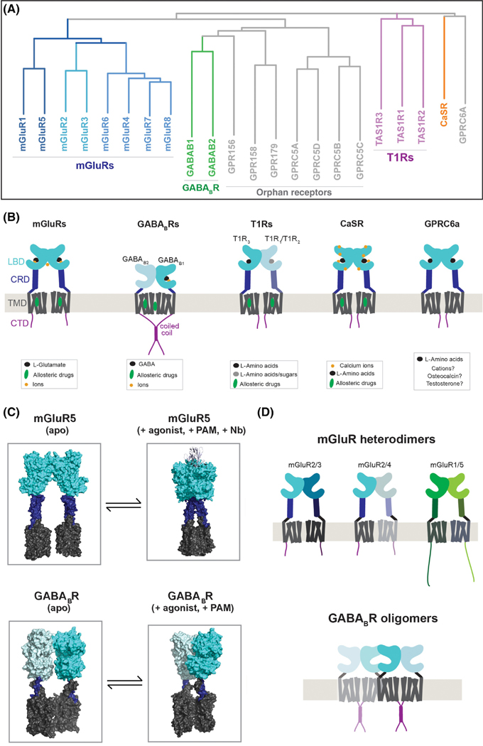
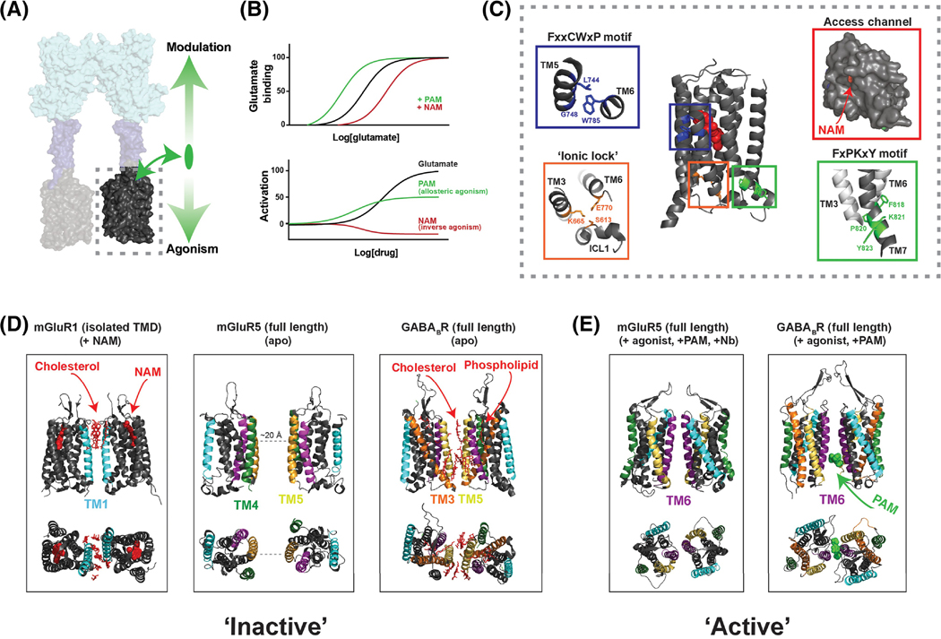
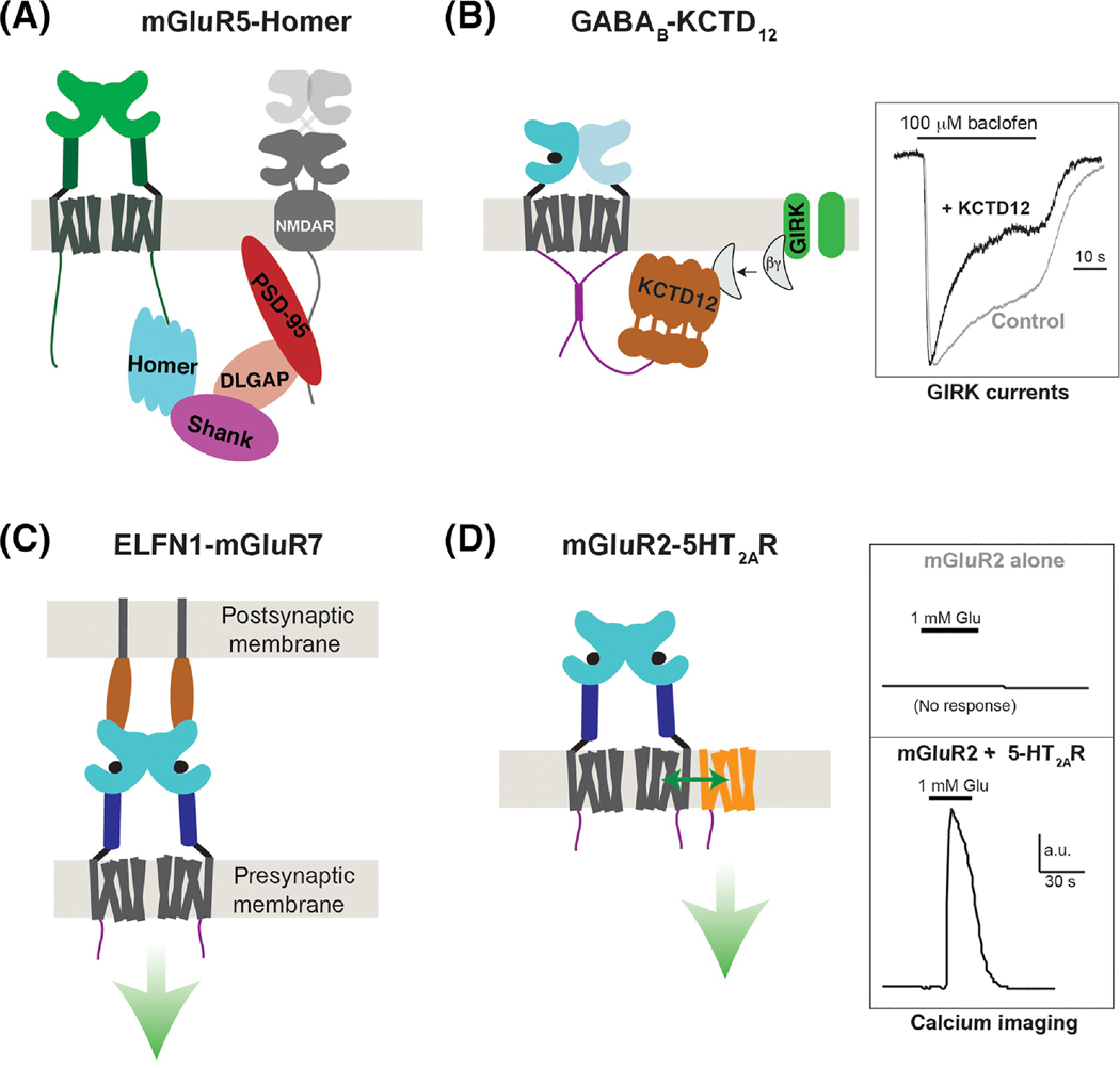
Groundbreaking structural and spectroscopic studies of class A G protein-coupled receptors (GPCRs), such as rhodopsin and the β2 adrenergic receptor, have provided a picture of how structural rearrangements between transmembrane helices control ligand binding, receptor activation, and effector coupling. However, the activation mechanism of other GPCR classes remains more elusive, in large part due to complexity in their domain assembly and quaternary structure. In this review, we focus on the class C GPCRs, which include metabotropic glutamate receptors (mGluRs) and gamma-aminobutyric acid B (GABAB) receptors (GABABRs) most prominently. We discuss the unique biophysical questions raised by the presence of large extracellular ligand-binding domains (LBDs) and constitutive homo/heterodimerization. Furthermore, we discuss how recent studies have begun to unravel how these fundamental class C GPCR features impact the processes of ligand binding, receptor activation, signal transduction, regulation by accessory proteins, and crosstalk with other GPCRs.

Keywords: G protein-coupled receptors; GABA(B) receptor; calcium-sensing receptor; metabotropic glutamate receptor.


Figures

Figure 1,
Figure 1,. Molecular diversity of class C GPCRs
A, Phylogenetic tree showing all class C GPCRs grouped into the major subfamilies. B, Summary of domain structure, homo- and hetero-dimerization and ligand binding properties of well-characterized, non-orphan class C GPCRs. T1R1/T1R3 heterodimers form the umami receptor and T1R2/T1R3 heterodimers form the sweet receptor. While T1R2, CaSR and GPRC6a show promiscuity with regard to L-amino acids, they prefer glutamate, tryptophan and basic amino acids, respectively. In the case of GPRC6a, controversy exists over whether osteocalcin and testosterone can bind and where their binding sites are and a defined cation binding site has not been proposed. C, Summary of cryo-EM structures of mGluR5 [20] and the GABABR [24] showing the apo-state (left) and an agonist and PAM-bound state (right). Note: an agonistic nanobody is shown in the agonist-bound mGluR5 structure. D, Schematic of further complexity in the assembly of class C GPCRs. Various heterodimeric mGluR combinations have been identified (top) and evidence for higher order assembly (bottom) exists, with the strongest data obtained for GABABRs. Oligomeric inter-LBD and inter-TMD interfaces involving GABAB1 subunits have been proposed for tetrameric or higher order GABAB complexes.
Figure 2,
Figure 2,. Structural dynamics of class C GPCR ligand binding domains (LBDs)
A, LBD dimers (dashed box) initiate activation following binding to orthosteric ligands, as shown in the full-length mGluR5 structure. B, Summary of mGluR LBD conformational changes. In an apo structure of mGluR1 (left) LBDs are found in the “open” state with upper (LB1) and lower (LB2) lobes far apart. At the inter-subunit level this structure is characterized as “relaxed” due to the lack of an inter-LBD interface. In a glutamate-bound structure of mGluR2 (right) both LBDs are in the closed state and dimer reorientation has allowed for the formation of an electrostatic LBD interface to form the “active state”. Note that the O-O/R and C-C/A are thought of as extreme conformations, that many intermediates exist and that the correlation between ligand occupancy and conformation is complex. C, 6-state model of mGluR LBD activation incorporating intra-subunit and inter-subunit conformational changes. * indicates states that have been captured in crystal structures. A 3-state model based on smFRET studies is highlighted with donor and acceptor fluorophore positions shown as green and red ovals. Right, representative smFRET trace showing the transition of mGluR2 between three states (dotted lines) on tens of ms time scale at approximately EC 50 glutamate levels. Inset shows the relative occupancy of the C-C/A state observed for mGluR2 under different conditions. D, Free energy diagrams summarizing differences in relative stability of relaxed and active states for mGluR2, 3 and 7. mGluR3 shows basal occupancy of the active state while mGluR7 shows minimal occupancy of the active state, even under saturating agonist conditions. E, GABABR LBD dimer structures showing a comparatively subtle dimer reorientation associated with activation.
Figure 3,
Figure 3,. Allosteric modulation and conformational dynamics of class C GPCR transmembrane domains
A, TMDs (dashed box) respond to agonist-binding in the LBD and mediate G protein activation. TMDs also bind allosteric ligands which can directly alter receptor activation (downward arrow) and modulate the response to orthosteric ligands (upward arrow). B, Theoretical dose-response curves showing the classical effects of allosteric modulators on orthosteric agonist binding (top) and on receptor activation (bottom). Many ligands show properties of both allosteric modulation and agonism or inverse agonism. C, NAM-bound mGluR5 TMD structure (PDB: 4OO9) reveals microswitches similar to those seen in class A structures, including the ionic lock interaction between Lys665 and Glu770 (orange box), the FxxCWxP motif (blue box) which is thought to serve as a “trigger switch” to couple ligand binding to TM6 rearrangement, and the FxPKxY motif (green box) at the intracellular end of TM7 which is thought to stabilize the active conformation. Entrance to the allosteric pocket is restricted by a narrow access channel formed by the helical bundle and extracellular loop 2 (red box). D-E, Structural data showing various inter-TMD dimer interfaces. NAM-bound mGluR1 TMD structures (D, left) revealed an inter-TM1 interface mediated, in part, by cholesterol molecules (red). The cryo-EM structure of mGluR5 showed no direct interface in the apo-state (D, center), but inactive GABABR cryo-EM structures show an interface consisting primarily of TM5 and the cytosolic end of TM3 as well as bound phospholipids and cholesterol (D, right)[23]. The agonist and PAM-bound cryo-EM structure of mGluR5 show an inter-TM6 interface (E, left) [24] that is similar to that seen in agonist and PAM-bound GABABR structures (E, right).
Figure 4,
Figure 4,. Modulation of class C GPCRs via accessory proteins
A, Schematic showing group I mGluR (mGluR1/5) coupling to a network of intracellular scaffold proteins which control signaling within the post-synaptic density. In brief, mGluR1 or mGluR5 CTDs bind directly to Homer proteins which, via SHANK, DLGAP and PSD-95, facilitate cross-talk with NMDA-type ionotropic glutamate receptors. Homer has also been shown to facilitate coupling to IP3 receptors and other elements of the scaffold and trafficking machinery. B, Interaction between the GABAB2 CTD and KCTD12 enhances the activation kinetics and desensitization of agonist-induced GIRK potassium channel currents by binding Gβγ subunits (right). Note: crystal structures revealed that the KCTD12 BTB domain forms a ring structure that binds one CTD and the H1 domain forms a 5:5 pentameric complex with Gβγ to effectively scavenge the G proteins away from the channel. C, Trans-synaptic interactions between group III mGluRs and ELFN proteins control the synaptic localization of mGluRs and have been shown to allosterically modulate receptor activation properties (green arrow), although the effects on signaling are unclear. D, Heteromerization between mGluR2 and class A 5-HT2ARs enables various forms of functional crosstalk, including trans-activation of 5-HT2ARs to produce Gq signaling (i.e. calcium elevation) following mGluR2 agonism (right). The stability and stoichiometry of such heteromers are not clear but an interface involving the cytoplasmic end of TM4 has been demonstrated.

References

    1. Sriram K and Insel PA (2018) G Protein-Coupled Receptors as Targets for Approved Drugs: How Many Targets and How Many Drugs? Mol Pharmacol 93 (4), 251–258. - PMC - PubMed
    1. de Graaf C. et al. (2017) Extending the Structural View of Class B GPCRs. Trends Biochem Sci 42 (12), 946–960. - PubMed
    1. Pin JP and Bettler B. (2016) Organization and functions of mGlu and GABAB receptor complexes. Nature 540 (7631), 60–68. - PubMed
    1. Kowatsch C. et al. (2019) Structures of vertebrate Patched and Smoothened reveal intimate links between cholesterol and Hedgehog signalling. Curr Opin Struct Biol 57, 204–214. - PMC - PubMed
    1. Zhang X. et al. (2018) Structural and Druggability Landscape of Frizzled G Protein-Coupled Receptors. Trends Biochem Sci 43 (12), 1033–1046. - PubMed

Publication types

MeSH terms

LinkOut - more resources