

Consistent localization of SARS-CoV-2 spike glycoprotein and ACE2 over TMPRSS2 predominance in placental villi of 15 COVID-19 positive maternal-fetal dyads
- PMID: 32862058
- PMCID: PMC7445146
- DOI: 10.1016/j.placenta.2020.08.015
Consistent localization of SARS-CoV-2 spike glycoprotein and ACE2 over TMPRSS2 predominance in placental villi of 15 COVID-19 positive maternal-fetal dyads
Abstract
Introduction: While the COVID-19 pandemic continues to have a significant global health impact, rates of maternal to infant vertical transmission remain low (<5%). Parenchymal changes of placentas from COVID-19 infected mothers have been reported by several groups, but the localization and relative abundance of SARS-CoV-2 viral proteins and cellular entry machinery has not been fully characterized within larger placental tissue cohorts.
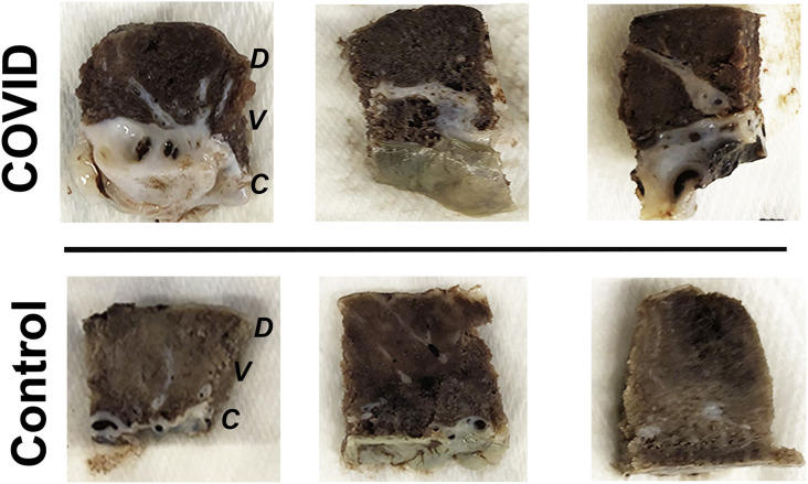
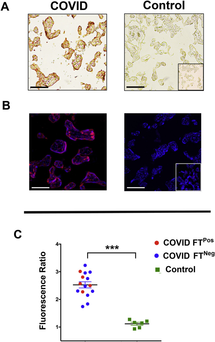
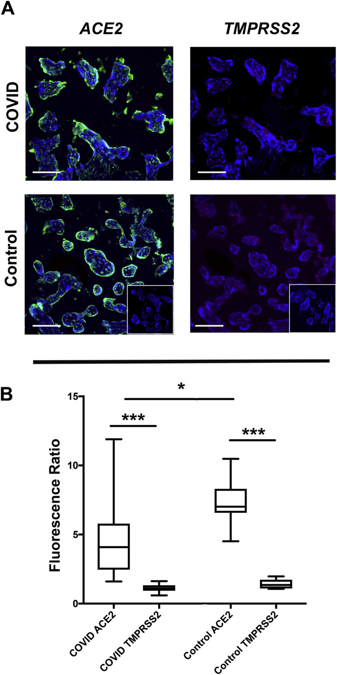
Methods: An extended placental tissue cohort including samples from 15 COVID-19 positive maternal-fetal dyads (with n = 5 cases with evidence of fetal transmission) in comparison with 10 contemporary COVID-19 negative controls. Using comparative immunofluorescence, we examined the localization and relative tissue abundance of SARS-CoV2 spike glycoprotein (CoV2 SP) along with the co-localization of two SARS-CoV2 viral entry proteins angiotensin-converting enzyme 2 (ACE2) and transmembrane serine protease 2 (TMPRSS2).
Results/conclusions: CoV2 SP was present within the villous placenta in COVID-19 positive pregnancies with and without evidence of fetal transmission. We further identified the predominance of ACE2 expression in comparison with TMPRSS2. Importantly, both CoV2 SP and ACE2 expression consistently localized primarily within the outer syncytiotrophoblast layer placental villi, a key physiologic interface between mother and fetus. Overall this study provides an important basis for the ongoing evaluation of SARS-CoV-2 physiology in pregnancy and highlights the importance of the placenta as a key source of primary human tissue for ongoing diagnostic and therapeutic research efforts to reduce the global burden of COVID-19.
Keywords: ACE2; COVID-19; Placenta; SARS-CoV-2; TMPRSS2; Vertical transmission.
Copyright © 2020 Elsevier Ltd. All rights reserved.
Conflict of interest statement
None.
Figures
Similar articles
-
SARS-CoV-2 can infect the placenta and is not associated with specific placental histopathology: a series of 19 placentas from COVID-19-positive mothers.Mod Pathol. 2020 Nov;33(11):2092-2103. doi: 10.1038/s41379-020-0639-4. Epub 2020 Aug 2. Mod Pathol. 2020. PMID: 32741970 Free PMC article.
-
Single-cell expression profiles of ACE2 and TMPRSS2 reveals potential vertical transmission and fetus infection of SARS-CoV-2.Aging (Albany NY). 2020 Oct 26;12(20):19880-19897. doi: 10.18632/aging.104015. Epub 2020 Oct 26. Aging (Albany NY). 2020. PMID: 33104520 Free PMC article.
-
Term Human Placental Trophoblasts Express SARS-CoV-2 Entry Factors ACE2, TMPRSS2, and Furin.mSphere. 2021 Apr 14;6(2):e00250-21. doi: 10.1128/mSphere.00250-21. mSphere. 2021. PMID: 33853873 Free PMC article.
-
Placental barrier against COVID-19.Placenta. 2020 Sep 15;99:45-49. doi: 10.1016/j.placenta.2020.07.022. Epub 2020 Jul 25. Placenta. 2020. PMID: 32755724 Free PMC article. Review.
-
Placental Pathology of COVID-19 with and without Fetal and Neonatal Infection: Trophoblast Necrosis and Chronic Histiocytic Intervillositis as Risk Factors for Transplacental Transmission of SARS-CoV-2.Viruses. 2020 Nov 15;12(11):1308. doi: 10.3390/v12111308. Viruses. 2020. PMID: 33203131 Free PMC article. Review.
Cited by
-
SARS-CoV-2 Infection and Placental Pathology.Rev Bras Ginecol Obstet. 2021 Jun;43(6):474-479. doi: 10.1055/s-0041-1730291. Epub 2021 Jun 2. Rev Bras Ginecol Obstet. 2021. PMID: 34077991 Free PMC article. Review.
-
Does COVID-19 cause pre-eclampsia?Ultrasound Obstet Gynecol. 2022 Feb;59(2):146-152. doi: 10.1002/uog.24809. Epub 2022 Jan 13. Ultrasound Obstet Gynecol. 2022. PMID: 34766403 Free PMC article. No abstract available.
-
Histopathological features in advanced abdominal pregnancies co-infected with SARS-CoV-2 and HIV-1 infections: A case evaluation.Eur J Obstet Gynecol Reprod Biol X. 2022 May 14;15:100153. doi: 10.1016/j.eurox.2022.100153. eCollection 2022 Aug. Eur J Obstet Gynecol Reprod Biol X. 2022. PMID: 35600136 Free PMC article.
-
Placental Infection Associated with SARS-CoV-2 Wildtype Variant and Variants of Concern.Viruses. 2023 Sep 13;15(9):1918. doi: 10.3390/v15091918. Viruses. 2023. PMID: 37766324 Free PMC article.
-
A standardized definition of placental infection by SARS-CoV-2, a consensus statement from the National Institutes of Health/Eunice Kennedy Shriver National Institute of Child Health and Human Development SARS-CoV-2 Placental Infection Workshop.Am J Obstet Gynecol. 2021 Dec;225(6):593.e1-593.e9. doi: 10.1016/j.ajog.2021.07.029. Epub 2021 Aug 5. Am J Obstet Gynecol. 2021. PMID: 34364845 Free PMC article.
References
-
- Map C.- Johns Hopkins Coronavirus Resource Center; 2020. COVID-19 Map.
-
- Yu N., Li W., Kang Q., Xiong Z., Wang S., Lin X., Liu Y., Xiao J., Liu H., Deng D., Chen S., Zeng W., Feng L., Wu J. Clinical features and obstetric and neonatal outcomes of pregnant patients with COVID-19 in Wuhan, China: a retrospective, single-centre, descriptive study, the Lancet. Infectious diseases. 2020;20(5):559–564. - PMC - PubMed
-
- Chen H., Guo J., Wang C., Luo F., Yu X., Zhang W., Li J., Zhao D., Xu D., Gong Q., Liao J., Yang H., Hou W., Zhang Y. Clinical characteristics and intrauterine vertical transmission potential of COVID-19 infection in nine pregnant women: a retrospective review of medical records. Lancet (London, England) 2020;395(10226):809–815. - PMC - PubMed
-
- Elshafeey F., Magdi R., Hindi N., Elshebiny M., Farrag N., Mahdy S., Sabbour M., Gebril S., Nasser M., Kamel M., Amir A., Maher Emara M., Nabhan A. A systematic scoping review of COVID-19 during pregnancy and childbirth. Int. J. Gynaecol. Obstet.: the official organ of the International Federation of Gynaecology and Obstetrics. 2020;150(1):47–52. - PMC - PubMed
Publication types
MeSH terms
Substances
Grants and funding
LinkOut - more resources
Full Text Sources
Miscellaneous
