Review
. 2020 Oct 30;295(44):15158-15171.
doi: 10.1074/jbc.REV120.012317. Epub 2020 Aug 29.

Structure, function, and inhibitor targeting of HIV-1 Nef-effector kinase complexes

Affiliations
Review

Structure, function, and inhibitor targeting of HIV-1 Nef-effector kinase complexes

Ryan P Staudt et al. J Biol Chem. .

Abstract

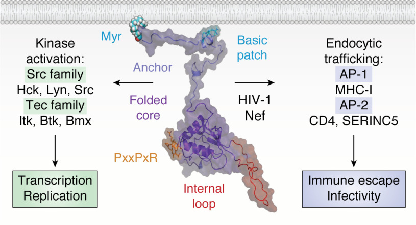
Antiretroviral therapy has revolutionized the treatment of AIDS, turning a deadly disease into a manageable chronic condition. Life-long treatment is required because existing drugs do not eradicate HIV-infected cells. The emergence of drug-resistant viral strains and uncertain vaccine prospects highlight the pressing need for new therapeutic approaches with the potential to clear the virus. The HIV-1 accessory protein Nef is essential for viral pathogenesis, making it a promising target for antiretroviral drug discovery. Nef enhances viral replication and promotes immune escape of HIV-infected cells but lacks intrinsic enzymatic activity. Instead, Nef works through diverse interactions with host cell proteins primarily related to kinase signaling pathways and endosomal trafficking. This review emphasizes the structure, function, and biological relevance of Nef interactions with host cell protein-tyrosine kinases in the broader context of Nef functions related to enhancement of the viral life cycle and immune escape. Drug discovery targeting Nef-mediated kinase activation has allowed identification of promising inhibitors of multiple Nef functions. Pharmacological inhibitors of Nef-induced MHC-I down-regulation restore the adaptive immune response to HIV-infected cells in vitro and have the potential to enhance immune recognition of latent viral reservoirs as part of a strategy for HIV clearance.

Keywords: AIDS; Btk; CD4; HIV-1 Nef; Hck; Itk; MHC-I; SH2 domain; SH3 domain; Src homology 2 domain (SH2 domain); Src homology 3 domain (SH3 domain); Src-family kinases; Tec-family kinases; bimolecular fluorescence complementation (BiFC); dimerization; endocytosis; human immunodeficiency virus (HIV); infectious disease; major histocompatibility complex (MHC); protein-protein interaction.


Conflict of interest statement

Conflict of interest—The authors declare that they have no conflicts of interest with the contents of this article.

Figures

Figure 1.
Figure 1.
Structural model of HIV-1 Nef and major classes of membrane-associated host cell partners. The overall structure of Nef is modeled in the center with major structural features indicated. The myristoylated (Myr) N-terminal anchor domain, together with a patch of basic amino acids from the amphipathic helix, tether Nef to the plasma membrane. The membrane anchor connects to the folded core, which encompasses multiple motifs essential for host cell protein recruitment. Highlighted is the conserved PxxPxR motif, which is essential for both SH3 domain interaction and binding to the AP-1 endocytic adaptor. Also shown is the flexible internal loop, which engages AP-2. Major classes of membrane-associated partner proteins include nonreceptor tyrosine kinases of the Src and Tec families (left), which Nef activates to enhance the viral life cycle. Nef also interacts with the endocytic adaptor proteins AP-1 and AP-2 to prevent cell-surface expression of multiple proteins, including MHC-I, CD4, and SERINC5, to promote viral infectivity and immune escape. This model of full-length Nef at the membrane is adapted from Geyer and Peterlin (19).
Figure 2.
Figure 2.
X-ray crystal structures of HIV-1 Nef in complex with the SH3 domain of the Src-family kinase, Fyn. A, overall structure of the Nef core in complex with the Fyn SH3 R96I mutant. Nef·SH3 complexes form a 2:2 dimer, with Nef forming the dimer interface. The Nef monomers are modeled in blue (NefA) and green (NefB), respectively, with the SH3 domains in red (SH3A) and pink (SH3B). B, SH3 domain surface residues Tyr-91, Trp-119, and Tyr-137 form hydrophobic grooves that contact the Nef PxxPxR motif (orange). This interaction is oriented and stabilized by a polar contact between SH3 Asp-100 and Arg-77 from the Nef PxxPxR motif. C, high-affinity Nef·SH3 interaction requires Ile-96 from the SH3 RT loop (red), which accesses a hydrophobic pocket created by Nef residues Phe-90, Trp-113, and Tyr-120 (cyan). Structural details of the Nef homodimer interface from this complex are shown in Fig. 5. Models were produced with PyMOL using the crystal coordinates of the HIV-1 Nef core in complex with the Fyn R96I mutant SH3 domain (PDB code 1EFN).
Figure 3.
Figure 3.
X-ray crystal structure of the HIV-1 Nef core in complex with the Hck SH3-SH2 regulatory region. The overall structure is shown on the left, which crystallized as a dimer of Nef·SH3-SH2 complexes. The surfaces of the Nef core monomers are rendered in blue (NefA) and green (NefB), respectively. The SH3-SH2 subunits associated with each Nef monomer are shown as ribbons, with SH3-SH2B in the foreground (SH2 in blue, SH3 in red). The SH3-SH2A subunit is in the background (SH2 in light blue, SH3 in pink) with the second SH3 domain partially hidden. A close-up view of one Nef interface is shown on the right, with the Nef PxxPxR motif (orange) contacting the SH3 surface via Pro-72 and Pro-75, with Arg-77 making an ionic contact with SH3 RT loop Asp-100. Unique to this complex is a second polar contact between SH3 RT loop Glu-94 and Arg-105 from the opposing Nef monomer. Details of the unique Nef homodimer interface found in this structure are highlighted in Fig. 6. Modeling was performed with PyMOL using the crystal coordinates of the HIV-1 Nef core in complex with the Hck SH3-SH2 domain (PDB code 4U5W).
Figure 4.
Figure 4.
Nef-mediated activation of Itk requires Nef homodimers. A, Nef recruits Itk to the cytoplasmic face of the plasma membrane and induces 2:2 Nef·Itk complex formation. Regulatory domain displacement and kinase domain juxtaposition induces constitutive kinase activation through trans-autophosphorylation. Dimerization-defective Nef mutants still interact with Itk at the membrane but form inactive 1:1 complexes. 3, Src homology 3 domain; 2, Src homology 2 domain; K, kinase domain; P, activation loop phosphorylation. B, activation of the T-cell receptor complex normally requires antigen-loaded MHC molecules from an antigen-presenting cell. The MHC-bound receptor then activates the Src-family kinase Lck, which is associated with the cytosolic tail of CD4. Lck phosphorylates and activates Itk, which in turn activates phospholipase Cγ (PLCγ) by direct phosphorylation. Phospholipase Cγ generates diacylglycerol (DAG) and inositol triphosphate (IP3) via hydrolysis of membrane phosphatidylinositol 4,5-bisphosphate (PIP2), leading to activation of protein kinase Cθ (PKCθ) and the calcium-dependent protein-serine/threonine phosphatase, calcineurin (Cal). Protein kinase Cθ promotes activation of NF-κB via the CARMA1/BCL10/MALT1 complex (not shown), whereas calcineurin dephosphorylates NFAT to drive nuclear localization. The NF-κB and NFAT transcription factors both enhance transcription of the integrated HIV-1 provirus early in the viral life cycle (91). Direct activation of Itk by Nef at the membrane downstream of the TCR may promote viral transcription through this pathway (see “Nef and Tec-family kinases”). LTR, long terminal repeat.
Figure 5.
Figure 5.
Nef homodimer interface from the X-ray crystal structure in complex with a Src-family kinase SH3 domain. Overview of the Nef dimer structure is shown in the top left, with the Nef monomers rendered in blue and green, respectively. The αB helices that form the dimer interface are highlighted (SH3 domains not shown for clarity). The αB helices are enlarged in the bottom left, illustrating the side chains of Leu-112, Tyr-115, and Phe-121 from each monomer, which form the hydrophobic core of the interface. Also shown are the reciprocal ionic contacts between Arg-105 and Asp-123. Both models are rotated 90° on the right. These models were produced with PyMOL using the crystal coordinates of the HIV-1 Nef core in complex with the Fyn SH3 domain R96I mutant (PDB code 1EFN). A model of the overall Nef·SH3 crystal structure is shown in Fig. 2A.
Figure 6.
Figure 6.
Homodimer interface from the X-ray crystal structure of Nef in complex with the Hck SH3-SH2 regulatory region. Three views of this Nef homodimer are shown on the left with the Nef monomers rendered in blue and green, respectively (SH3-SH2 proteins not shown for clarity). Three interfaces stabilize this homodimer, which are enlarged on the right. In the top view, the side chains of Leu-112, Tyr-115, Phe-121, and Pro-122 form a hydrophobic cup that interacts with Val-70 from the opposing monomer in reciprocal fashion. The side view shows the contributions of Ile-109, Leu-112, Trp-113, and His-116 to a hydrophobic interface between the αB helices. The bottom view shows reciprocal polar contacts formed by Ser-103 and Arg-106 with the main-chain carbonyls of Gly-95 and Gly-96; Leu-100 also makes a nonpolar contact in this interface. These models were produced with PyMOL using the crystal coordinates of the HIV-1 Nef core in complex with the Hck SH3-SH2 region (PDB code 4U5W). A model of the overall Nef·SH3-SH2 crystal structure is shown in Fig. 3A.
Figure 7.
Figure 7.
Interaction of HIV-1 Nef with the Src-family kinase Hck may induce conformational changes consistent with MHC-I/AP-1 recruitment. Left, crystal structure of the HIV-1 Nef core in complex with the SH3 domain of Fyn (high-affinity R96I mutant; PDB code 1EFN). Nef forms a dimer of Nef·SH3 complexes in this structure (NefA (blue) and NefB (green)). The dimer interface is formed by the Nef αB helices (highlighted helices). The SH3 domains are shown at the top in pink. The helical dimer interface is enlarged in the bottom panel and highlights the reciprocal polar contacts between Nef Asp-123 and Arg-105, which are buried in the core of this structure. The center, top panel shows the crystal structure of the Nef core in complex with the Hck SH3-SH2 region (PDB code 4U5W). The color scheme is as per the left panel with a single SH3-SH2 unit shown for clarity. Nef also crystallizes as a 2:2 complex of Nef·SH3-SH2 dimers in this structure, but the helical interface is completely reoriented relative to the Nef·SH3 complex such that the Nef Asp-123 side chain (orange) is now pointed toward the solvent. One of the Asp-123 residues is enlarged in the bottom panel. Right, structural alignment of one of the Nef core proteins (green ribbon) in the Nef·SH3-SH2 complex (PDB code 4U5W) with the Nef core (cyan ribbon) in complex with the AP-1/μ1 subunit (pink) and MHC-I tail peptide (purple) from PDB code 4EN2. The Nef core proteins from each complex adopt very similar conformations, with nearly identical positioning of Nef Asp-123 (enlarged in the bottom panel). Nef Asp-123 forms part of an ionic bond network, with Arg-393 from AP-1 and Asp-327 from MHC-I, that is required for Nef-mediated down-regulation of MHC-I and immune escape.
Figure 8.
Figure 8.
Examples of small molecule inhibitors discovered using a Nef-coupled Hck kinase activity assay. Combining recombinant Nef and Hck proteins in vitro enabled high-throughput screening for inhibitors of the active complex (illustrated at the right; 3, SH3 domain; 2, SH2 domain; K, kinase domain). Screening of a small kinase inhibitor–biased library identified diphenylfuranopyrimidine 4-amino propanol (DFP-4AP), which inhibits the Nef-Hck complex via the Hck kinase domain (103). Screening of a large, more diverse library identified the compound B9 (102), which binds directly to Nef and inhibits Hck by an allosteric mechanism that may be related to Nef homodimer formation (left). Medicinal chemistry optimization has led to more potent analogs, such as FC-8052, which shares a hydroxypyrazole core with B9 (red). FC-8052 binds to recombinant Nef in vitro with a KD value of ∼10 pm, compared with about 80 nm for B9, and inhibits Nef-dependent HIV-1 replication in PBMCs in the subnanomolar range (100).

References

    1. Jaffe H. W. (2008) The early days of the HIV-AIDS epidemic in the U.S.A. Nat. Immunol. 9, 1201–1203 10.1038/ni1108-1201 - DOI - PubMed
    1. Marwick C. (1987) AZT (zidovudine) just a step away from FDA approval for AIDS therapy. JAMA 257, 1281–1282 10.1001/jama.1987.03390100015002 - DOI - PubMed
    1. Hammer S. M., Katzenstein D. A., Hughes M. D., Gundacker H., Schooley R. T., Haubrich R. H., Henry W. K., Lederman M. M., Phair J. P., Niu M., Hirsch M. S., and Merigan T. C. (1996) A trial comparing nucleoside monotherapy with combination therapy in HIV-infected adults with CD4 cell counts from 200 to 500 per cubic millimeter. AIDS Clinical Trials Group Study 175 Study Team. N. Engl. J. Med. 335, 1081–1090 10.1056/NEJM199610103351501 - DOI - PubMed
    1. Vella S., Schwartlander B., Sow S. P., Eholie S. P., and Murphy R. L. (2012) The history of antiretroviral therapy and of its implementation in resource-limited areas of the world. AIDS 26, 1231–1241 10.1097/QAD.0b013e32835521a3 - DOI - PubMed
    1. Sepkowitz K. A. (2001) AIDS–the first 20 years. N. Engl. J. Med. 344, 1764–1772 10.1056/NEJM200106073442306 - DOI - PubMed

Publication types

MeSH terms