. 2021 Feb;31(2):1081-1089.
doi: 10.1007/s00330-020-07126-8. Epub 2020 Aug 29.

Observer agreement and clinical significance of chest CT reporting in patients suspected of COVID-19

Affiliations

Observer agreement and clinical significance of chest CT reporting in patients suspected of COVID-19

Marie-Pierre Debray et al. Eur Radiol. 2021 Feb.

Abstract

Objectives: To assess interobserver agreement and clinical significance of chest CT reporting in patients suspected of COVID-19.

Methods: From 16 to 24 March 2020, 241 consecutive patients addressed to hospital for COVID-19 suspicion had both chest CT and SARS-CoV-2 RT-PCR. Eight observers (2 thoracic and 2 general senior radiologists, 2 junior radiologists, and 2 emergency physicians) retrospectively categorized each CT into one out of 4 categories (evocative, compatible for COVID-19 pneumonia, not evocative, and normal). Observer agreement for categorization between all readers and pairs of readers with similar experience was evaluated with the Kappa coefficient. The results of a consensus categorization were correlated to RT-PCR.

Results: Observer agreement across the 4 categories was good between all readers (κ value 0.61 95% CI 0.60-0.63) and moderate to good between pairs of readers (0.54-0.75). It was very good (κ 0.81 95% CI 0.79-0.83), fair (κ 0.32 95% CI 0.29-0.34), moderate (κ 0.56 95% CI 0.54-0.58), and moderate (0.58 95% CI 0.56-0.61) for the categories evocative, compatible, not evocative, and normal, respectively. RT-PCR was positive in 97%, 50%, 31%, and 11% of cases in the respective categories. Observer agreement was lower (p < 0.001) and RT-PCR positive cases less frequently categorized evocative in the presence of an underlying pulmonary disease (p < 0.001).

Conclusion: Interobserver agreement for chest CT reporting using categorization of findings is good in patients suspected of COVID-19. Among patients considered for hospitalization in an epidemic context, CT categorized evocative is highly predictive of COVID-19, whereas the predictive value of CT decreases between the categories compatible and not evocative.

Key points: • In patients suspected of COVID-19, interobserver agreement for chest CT reporting into categories is good, and very good to categorize CT "evocative." • Chest CT can participate in estimating the likelihood of COVID-19 in patients presenting to hospital during the outbreak, CT categorized "evocative" being highly predictive of the disease whereas almost a third of patients with CT "not evocative" had a positive RT-PCR in our study. • Observer agreement is lower and CTs of positive RT-PCR cases less frequently "evocative" in presence of an underlying pulmonary disease.

Keywords: Coronavirus infections; Pneumonia; Tomography, X-ray computed.


Conflict of interest statement

The authors of this manuscript declare no relationships with any companies whose products or services may be related to the subject matter of the article.

Figures

Fig. 1
Fig. 1
Flow chart of the study. SARS-CoV-2 RT PCR and chest CT were performed at presentation, except in 7 cases for which RT-PCR had been performed 1 to 3 days earlier. Among 158 COVID-19 confirmed cases, RT-PCR was positive at admission in 151 cases and in subsequent days during hospitalization in 7 cases.*Clinical and chest CT follow-up. **Absence of follow-up available because the patient had been transferred to another hospital or turned back home
Fig. 2
Fig. 2
Chest CT scan categorized “evocative of COVID-19 pneumonia” in two different patients with positive nasopharyngeal SARS-CoV-2 RT-PCR at admission and secondarly (panels a and b and panels c and e, respectively). Multifocal bilateral ground-glass opacities with subpleural and posterior predominance, associated with band-like (panel b) or more extensive consolidations (panel d)
Fig. 3
Fig. 3
Chest CT scan categorized “compatible with COVID-19 pneumonia,” in association with fibrosing interstitial lung disease (ILD) showing a non-specific interstitial pneumonia pattern (panel c, axial plane through the lung bases). Pure ground-glass opacities, both centrally and peripherally distributed, have appeared in the left upper lobe (panels a, b), as compared to the previous CT performed 4 months earlier (panel d). Such new opacities could be attributable to COVID-19, another infection, or acutisation of ILD. Nasopharyngeal SARS-CoV-2 RT-PCR positive
Fig. 4
Fig. 4
Chest CT scan categorized “compatible with with COVID-19 pneumonia,” in association with pulmonary edema, manifesting as ground-glass opacities with a predominant central distribution, septal lines, and bilateral pleural effusion (panels a, b, axial plane; panel c, coronal plane) in a patient with history of chronic renal insufficiency on dialysis. Subpleural consolidation (panel a) in the posterior zone of the right upper lobe is consistent with associated COVID-19 pneumonia. Nasopharyngeal SARS-CoV-2 RT-PCR positive
Fig. 5
Fig. 5
Chest CT scan categorized “not evocative of COVID-19 peumonia,” showing small consolidations in the left lower lobe associated with bronchial thickening and endobronchial filling, in favor of bronchopneumonia. Nasopharyngeal SARS-CoV-2 RT-PCR negative
Fig. 6
Fig. 6
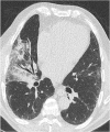
Chest CT scan categorized “not evocative of COVID-19 pneumonia” showing combination of areas of ground-glass opacity and consolidation systematized in the middle lobe (arrows), evoquing a lobar pneumonia. Despite positivity of the SARS-CoV-2-RT-PCR, bacterial co-infection was suspected and the patient received antibiotics with favorable evolution

Similar articles

Cited by

References

    1. Huang C, Wang Y, Li X, et al. Clinical features of patients infected with 2019 novel coronavirus in Wuhan, China. Lancet. 2020;395(10223):497–506. doi: 10.1016/S0140-6736(20)30183-5. - DOI - PMC - PubMed
    1. World Health Organization (2020) Director General’s speeches. https://www.who.int/dg/speeches/detail/who-director-general-s-opening-re.... Accessed 27 Apr 2020
    1. Sverzellati N, Milone F, Balbi M. How imaging should properly be used in COVID-19 outbreak: an Italian experience. Diagn Interv Radiol. 2020;26:204–206. doi: 10.5152/dir.2020.30320. - DOI - PMC - PubMed
    1. Ai T, Yang Z, Hou H et al (2020) Correlation of chest CT and RT-PCR testing in coronavirus disease 2019 (COVID-19) in China: a report of 1014 cases. Radiology. 10.1148/radiol.2020200642 - PMC - PubMed
    1. Fang Y, Zhang H, Xie J, et al. Sensitivity of chest CT for COVID-19: comparison to RT-PCR. Radiology. 2020;295:208–209. doi: 10.1148/radiol.2020200280. - DOI - PMC - PubMed