Review
. 2021 Jan;37(1):343-358.
doi: 10.1007/s10554-020-01980-9. Epub 2020 Aug 30.

Acute pulmonary embolism multimodality imaging prior to endovascular therapy

Affiliations
Review

Acute pulmonary embolism multimodality imaging prior to endovascular therapy

David Sin et al. Int J Cardiovasc Imaging. 2021 Jan.

Abstract

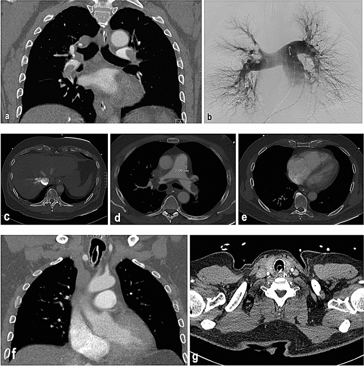
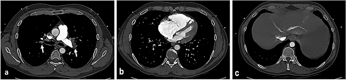
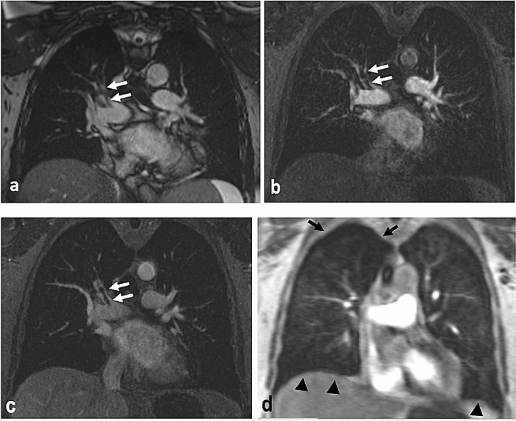
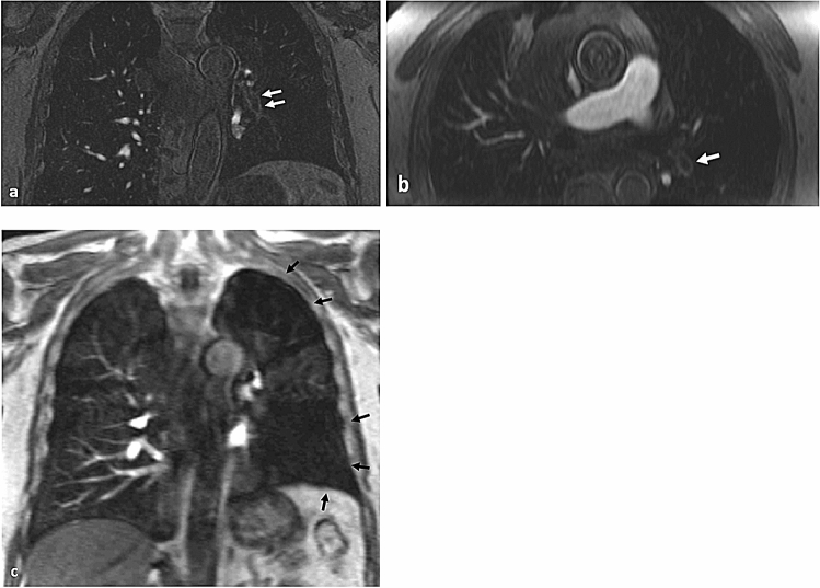
The manuscript discusses the application of CT pulmonary angiography, ventilation-perfusion scan, and magnetic resonance angiography to detect acute pulmonary embolism and to plan endovascular therapy. CT pulmonary angiography offers high accuracy, speed of acquisition, and widespread availability when applied to acute pulmonary embolism detection. This imaging modality also aids the planning of endovascular therapy by visualizing the number and distribution of emboli, determining ideal intra-procedural catheter position for treatment, and signs of right heart strain. Ventilation-perfusion scan and magnetic resonance angiography with and without contrast enhancement can also aid in the detection and pre-procedural planning of endovascular therapy in patients who are not candidates for CT pulmonary angiography.

Keywords: Acute pulmonary embolism; Computed tomography pulmonary angiography; Magnetic resonance angiography; Ventilation–perfusion scan.


Conflict of interest statement

The authors declares that they have no conflict of interest.

Figures

Fig. 1
Fig. 1
80-year-old female with known ANCA-negative, medium-size-vessel vasculitis presenting with progressive dyspnea over a period of 4 weeks. a Dual-energy CTPA demonstrates filling defects of several subsegmental arteries, one of them illustrated in this axial plane (arrow). b Z-effective map of the dual-energy CTPA demonstrating iodine distribution with blue colors representing high iodine concentration and yellow and red colors representing low iodine concentration. This axial plane at the same level shows a wedge-shaped area of low iodine concentration (arrows) corresponding to an area of reduced perfusion caused by the embolus seen in a. Further perfusion defects can be appreciated on the same plane (arrowheads) corresponding to more emboli not detected with regular CTPA imaging
Fig. 2
Fig. 2
64-year-old male with hepatocellular carcinoma. a Dual-energy CTPA demonstrates filling defects of several subsegmental arteries, one of them illustrated in this axial plane (arrow). b Z-effective map of the dual-energy CTPA illustrates a wedge-shaped area of low iodine concentration at the same level corresponding to an area of reduced perfusion (arrow)
Fig. 3
Fig. 3
69-year-old male with a massive pulmonary embolism status post cardiac arrest with ROSC achieved after CPR. a Coronal view CTPA showing bilateral pulmonary artery emboli extending into segmental branches. b Digital subtraction angiography of pulmonary arteries via right common femoral vein access with 5 Fr flush pigtail drainage catheter in main pulmonary artery showing bilateral acute PE, predominantly on the right side. Correlation between coronal CTPA and DSA images is helpful to determine extend of disease. ce Signs of right heart strain and acute PE on axial CTPA imaging including reflux of contrast into hepatic veins and IVC (c), acute dilatation of main pulmonary artery (d) as well as RV to LA ratio of more than 1 with straightening of the interventricular septum. f and g are representative coronal and axial CTPA slices showing patent SVC and patent bilateral internal jugular veins. This information can be gained from the CTPA and is helpful for procedure planning purposes, particularly if the endovascular treatment approach will be pursued via internal jugular vein access
Fig. 4
Fig. 4
48-year-old female presenting with severe shortness of breath and chest pain. The patient has a history of metastatic breast cancer. ac Coronal CTPA slices showing pulmonary embolus in right and left pulmonary arteries extending into multiple segmental branches. d RV to LV of 1.0 suggestive of right heart strain. e In this case there is no significant contrast reflux into suprahepatic inferior vena cava and / or hepatic veins
Fig. 5
Fig. 5
45-year-old male patient with acute dyspnea. a CTPA shows emboli in the left pulmonary artery bifurcation and the lower lobe segmental artery on the right side (arrows) b Significant enlargement of the right ventricle with a nearly inversed configuration of the interventricular septum, consistent with right heart strain. c A less specific, but also typical sign of right heart strain is the reflux of contrast material into the inferior vena cava and hepatic veins
Fig. 6
Fig. 6
62-year-old female patient with acute dyspnea. a Static SSFP sequence acquired in coronal orientation shows a filling defect in the right upper lobe artery (white arrows). b and c Contrast-enhanced 3D MRA acquired in coronal orientation in the pulmonary arterial phase (b) and 25 s later (c) confirms the findings of the SSFP sequence (white arrows). d Time-resolved, contrast-enhanced 3D MRA demonstrates a corresponding perfusion defect in the right upper lobe (black arrows) as well as further perfusion defects in the right and left basal segments (black arrow heads, corresponding thromboemboli not illustrated in this figure)
Fig. 7
Fig. 7
69-year-old male with acute dyspnea. a and b Contrast-enhanced 3D MRA acquired in coronal orientation demonstrates filling defects, among others a long filling defect in the left lower lobe artery with a “railway sign” (a, white arrows) and the filling defect is shown as a “polo mint sign” on the axial reconstruction of the same data set (b, white arrow). c Time-resolved, contrast-enhanced 3D MRA reveals extensive wedge-shaped perfusion defects in the left upper and lower lobes (black arrows)

References

    1. Raja AS, Greenberg JO, Qaseem A, Denberg TD, Fitterman N, Schuur JD, for the Clinical Guidelines Committee of the American College of Physicians Evaluation of patients with suspected acute pulmonary embolism: best practice advice from the Clinical Guidelines Committee of the American College of Physicians. Ann Intern Med. 2015;163:701. doi: 10.7326/M14-1772. - DOI - PubMed
    1. Konstantinides SV, Torbicki A, Agnelli G, Danchin N, Fitzmaurice D, Galiè N, Gibbs JSR, Huisman MV, Humbert M, Kucher N, Lang I, Lankeit M, Lekakis J, Maack C, Mayer E, Meneveau N, Perrier A, Pruszczyk P, Rasmussen LH, Schindler TH, Svitil P, Vonk Noordegraaf A, Zamorano JL, Zompatori M, Task Force for the Diagnosis and Management of Acute Pulmonary Embolism of the European Society of Cardiology (ESC) 2014 ESC guidelines on the diagnosis and management of acute pulmonary embolism. Eur Heart J. 2014;35:3033–3069. doi: 10.1093/eurheartj/ehu283. - DOI - PubMed
    1. Alotaibi GS, Wu C, Senthilselvan A, McMurtry MS. Secular trends in incidence and mortality of acute venous thromboembolism: the AB-VTE Population-Based Study. Am J Med. 2016;129:879.e19–25. doi: 10.1016/j.amjmed.2016.01.041. - DOI - PubMed
    1. Horlander KT, Mannino DM, Leeper KV. Pulmonary embolism mortality in the United States, 1979–1998: an analysis using multiple-cause mortality data. Arch Intern Med. 2003;163:1711–1717. doi: 10.1001/archinte.163.14.1711. - DOI - PubMed
    1. Silverstein MD, Heit JA, Mohr DN, Petterson TM, O’Fallon WM, Melton LJ. Trends in the incidence of deep vein thrombosis and pulmonary embolism: a 25-year population-based study. Arch Intern Med. 1998;158:585–593. doi: 10.1001/archinte.158.6.585. - DOI - PubMed

MeSH terms