. 2020 Oct;76(4):1195-1207.
doi: 10.1161/HYPERTENSIONAHA.120.15384. Epub 2020 Aug 31.

Altered Cardiovascular Defense to Hypotensive Stress in the Chronically Hypoxic Fetus

Affiliations

Altered Cardiovascular Defense to Hypotensive Stress in the Chronically Hypoxic Fetus

Beth J Allison et al. Hypertension. 2020 Oct.

Abstract

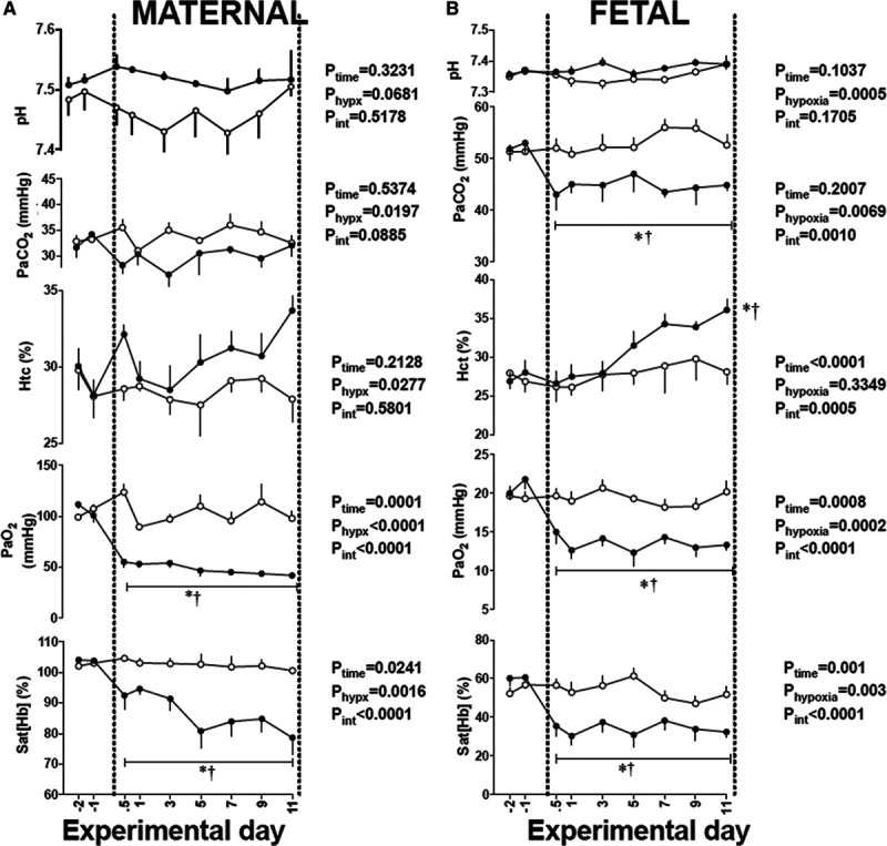
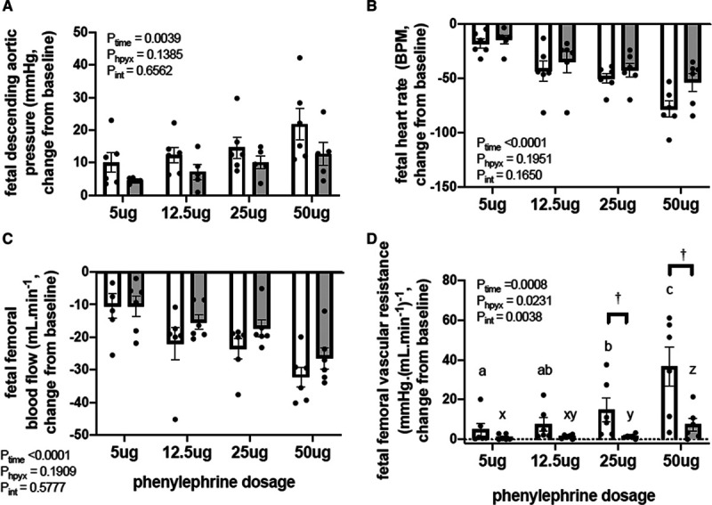
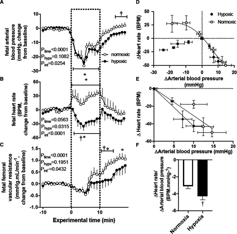
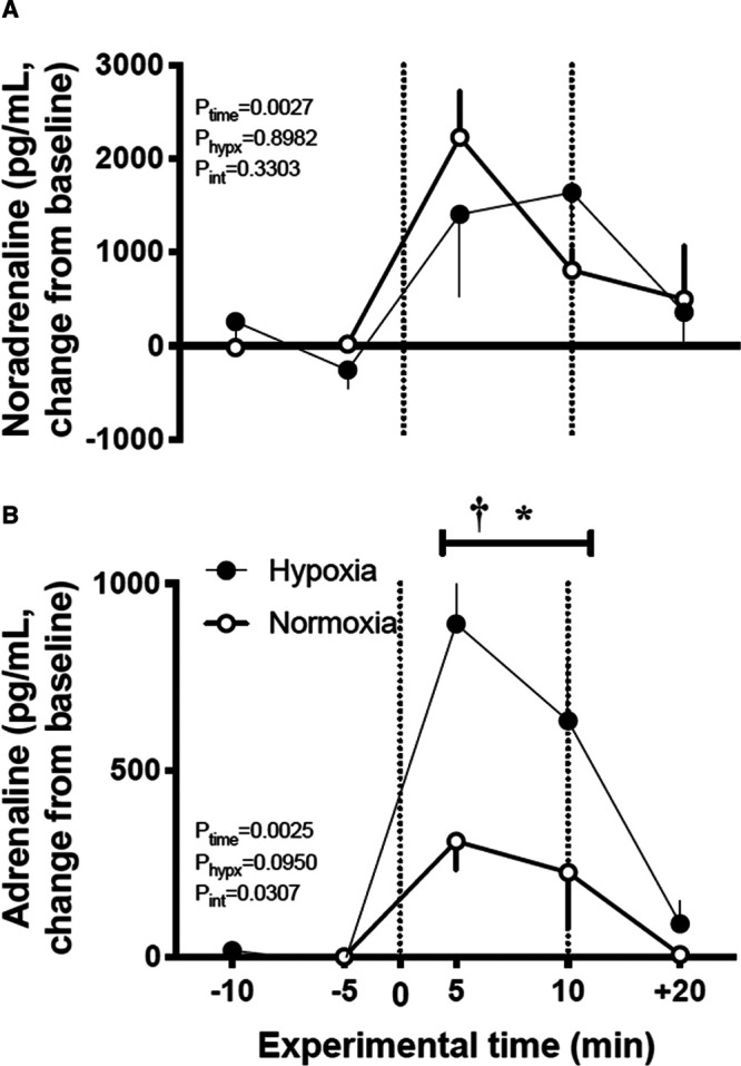
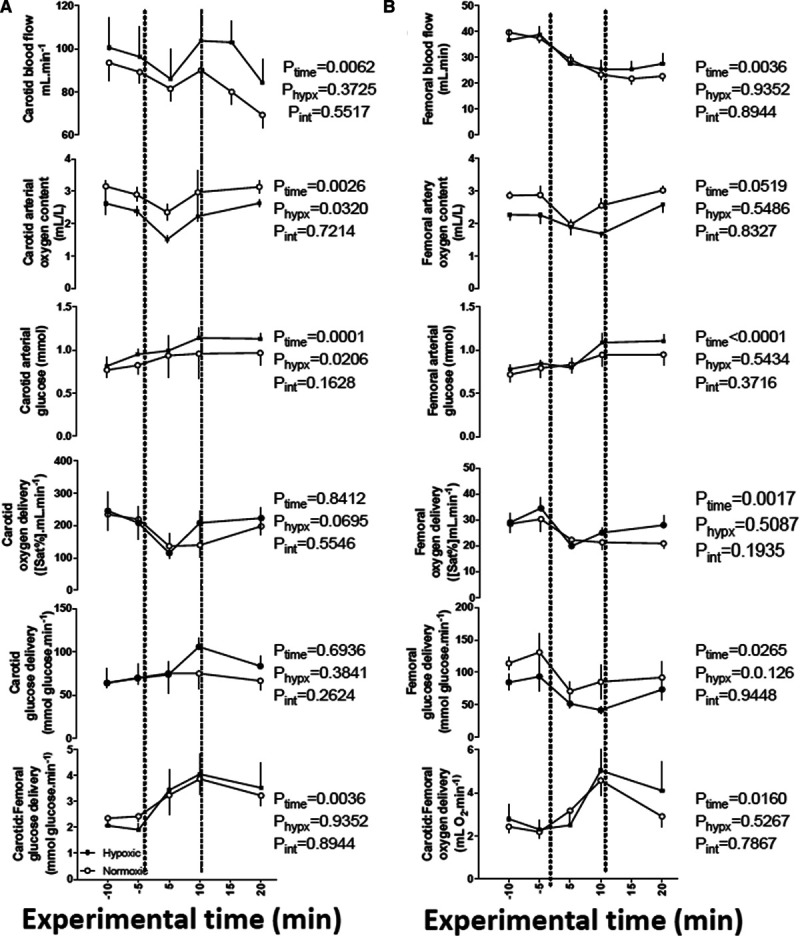
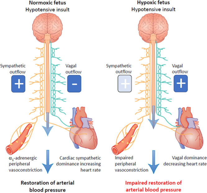
The hypoxic fetus is at greater risk of cardiovascular demise during a challenge, but the reasons behind this are unknown. Clinically, progress has been hampered by the inability to study the human fetus non-invasively for long period of gestation. Using experimental animals, there has also been an inability to induce gestational hypoxia while recording fetal cardiovascular function as the hypoxic pregnancy is occurring. We use novel technology in sheep pregnancy that combines induction of controlled chronic hypoxia with simultaneous, wireless recording of blood pressure and blood flow signals from the fetus. Here, we investigated the cardiovascular defense of the hypoxic fetus to superimposed acute hypotension. Pregnant ewes carrying singleton fetuses surgically prepared with catheters and flow probes were randomly exposed to normoxia or chronic hypoxia from 121±1 days of gestation (term ≈145 days). After 10 days of exposure, fetuses were subjected to acute hypotension via fetal nitroprusside intravenous infusion. Underlying in vivo mechanisms were explored by (1) analyzing fetal cardiac and peripheral vasomotor baroreflex function; (2) measuring the fetal plasma catecholamines; and (3) establishing fetal femoral vasoconstrictor responses to the α1-adrenergic agonist phenylephrine. Relative to controls, chronically hypoxic fetal sheep had reversed cardiac and impaired vasomotor baroreflex function, despite similar noradrenaline and greater adrenaline increments in plasma during hypotension. Chronic hypoxia markedly diminished the fetal vasopressor responses to phenylephrine. Therefore, we show that the chronically hypoxic fetus displays markedly different cardiovascular responses to acute hypotension, providing in vivo evidence of mechanisms linking its greater susceptibility to superimposed stress.

Keywords: blood pressure; fetus; hypotension; pregnancy; sheep.


Conflict of interest statement

License agreement 100395 CamDAS: Technology for simultaneous wireless recording of arterial blood pressure and blood flow in large animals. D.A. Giussani, Maastricht Instruments, The British Heart Foundation and Cambridge Enterprise. The other authors report no conflicts.

Figures

Figure 1.
Figure 1.
Maternal and fetal blood gases during chronic hypoxia. Values are mean±SEM. for the maternal (A) and fetal (B) arterial blood gas status in sheep undergoing normoxic (○, n=6) or chronic hypoxic (•, n=6) pregnancy. The results of the 2-way RM ANOVA for main effects and interactions are shown. When a significant (P<0.05) interaction between main effects occurred, differences were compared using the Tukey post hoc test. * indicates a significant effect of time compared with baseline; † indicates a significant effect of treatment compared with normoxic pregnancy. Htc indicates hematocrit; PaO2, arterial O2 partial pressure; PaCO2, arterial CO2 partial pressure; pH, arterial pH; and Sat [Hb], percentage saturation of hemoglobin.
Figure 2.
Figure 2.
Pressor and femoral vasopressor responses to phenylephrine in the chronically hypoxic fetus. Values are the mean±SEM for the change from baseline in fetal arterial blood pressure (A), heart rate (B), femoral blood flow (C), and femoral vascular resistance (D) in response to increasing doses of phenylephrine (5, 12.5, 25, and 50 μg) in normoxic (open bars, n=6) and chronically hypoxic (gray bars, n=6) fetuses. The results of the 2-way RM ANOVA for main effects and interactions are shown. When a significant (P<0.05) interaction between main effects occurred, differences were compared using the Newman-Keuls post hoc test. Different letters indicate a significant effect of dose within groups (normoxic a–c, hypoxic x–z); † indicates a significant effect of treatment compared with normoxic pregnancy.
Figure 3.
Figure 3.
Cardiovascular responses to acute hypotension and cardiac baroreflex function in the chronically hypoxic fetus. A–C, Show the mean±SEM values for the change from baseline in arterial blood pressure (A), heart rate (B), and femoral vascular resistance (C) in normoxic (○, n=6) or chronically hypoxic (•, n=6) fetuses during the acute hypotension experiment. Acute hypotension (dashed box) was induced for 10 min by fetal intravenous infusion with sodium nitroprusside (2.5 mg/kg per min). D–F, Show an analysis of cardiac baroreflex function. The fetal arterial blood pressure-heart rate relationship was constructed by plotting the change from baseline in both variables mean±SEM (x and y) in response to sodium nitroprusside or to fetal treatment with increasing doses of phenylephrine (D). The vagal dominant component of the cardiac baroreflex is expanded in (E; mean±SEM. for x and y). F, Shows the mean±SEM values for an analysis of slopes for the vagal dominant component of the cardiac baroreflex. The results of the 2-way RM ANOVA for main effects and interactions for A–C are shown. When a significant (P<0.05) interaction between main effects occurred, differences were compared using the Tukey post hoc test. Groups in (F) were compared by the Student t test for unpaired data. *indicates a significant effect of time compared with baseline; †indicates a significant effect of treatment compared with normoxic pregnancy.
Figure 4.
Figure 4.
Plasma catecholamine response to acute hypotension in the chronically hypoxic fetus. Values are mean±SEM for the change from baseline in noradrenaline (A) and adrenaline (B) in normoxic (○, n=6) or chronically hypoxic (•, n=6) fetuses during the acute hypotension experiment. Acute hypotension (dashed box) was induced for 10 min by fetal intravenous infusion with sodium nitroprusside (2.5 mg/kg per min). Plasma samples were taken during baseline (−10 and −5 min), during hypotension (+5 and +10 min) and after 10 min of recovery (+20 min). The results of the 2-way RM ANOVA for main effects and interactions are shown. When a significant (P<0.05) interaction between main effects occurred, differences were compared using the Tukey post hoc test. *indicates a significant effect of time compared with baseline; †indicates a significant effect of treatment compared with normoxic pregnancy.
Figure 5.
Figure 5.
Changes in oxygen and glucose delivery in the carotid and femoral vascular beds during acute hypotension in the chronically hypoxic fetus. Values are mean±SEM for blood flow, oxygen content, blood glucose concentration, oxygen delivery, and glucose delivery in the carotid (A) and femoral (B) vascular beds. The ratio of oxygen and glucose delivery in the carotid relative to the femoral vascular beds is also calculated. Groups are normoxic (○, n=6) or chronically hypoxic (•, n=6) fetuses. Acute hypotension (dashed box) was induced for 10 min by fetal intravenous infusion with sodium nitroprusside (2.5 mg/kg per min). The results of the 2-way RM ANOVA for main effects and interactions are shown. There are no significant interactions between main effects.
Figure 6.
Figure 6.
Summary figure. During acute hypotension, the normoxic fetus in healthy pregnancy activates cardiac and vasomotor baroreflex responses to restore blood pressure homeostasis. This involves baroreflex alteration in autonomic outflow via the brain stem. Increased sympathetic outflow coupled with vagal withdrawal to the heart promotes cardiac sympathetic dominant effects, which increase heart rate and contractility via β1-adrenergic receptor pathways. Enhanced sympathetic outflow to the arterioles increases peripheral vascular resistance via a1-adrenergic receptor pathways. Enhanced sympathetic outflow to the veins increases venous return via a1-adrenergic receptor pathways. The increase in venous return, coupled with increased myocardial contractility lead to an increase in cardiac output. The increase in peripheral vascular resistance together with the increase in cardiac output restores fetal arterial blood pressure back to baseline. In the chronically hypoxic fetus, baroreflex-induced increases in heart rate do not occur. Instead, there is a fall in heart rate because of increased vagal dominant effects. In addition, responsiveness to α1-adrenergic receptor pathway stimulation is blunted in the peripheral vasculature. Combined, these effects of chronic hypoxia on the heart and circulation weaken the fetal cardiovascular defense to acute hypotension in the chronically hypoxic fetus, providing a mechanistic link underlying its greater susceptibility to a second hit.

Similar articles

Cited by

References

    1. Hutter D, Kingdom J, Jaeggi E. Causes and mechanisms of intrauterine hypoxia and its impact on the fetal cardiovascular system: a review. Int J Pediatr. 2010;2010:401323. doi: 10.1155/2010/401323. - PMC - PubMed
    1. Rudolph AM, Itskovitz J, Iwamoto H, Reuss ML, Heymann MA. Fetal cardiovascular responses to stress. Semin Perinatol 19815109–121 - PubMed
    1. Giussani DA. The fetal brain sparing response to hypoxia: physiological mechanisms. J Physiol 20165941215–1230doi: 10.1113/JP271099 - PMC - PubMed
    1. Ducsay CA, Goyal R, Pearce WJ, Wilson S, Hu XQ, Zhang L. Gestational hypoxia and developmental plasticity. Physiol Rev 2018981241–1334doi: 10.1152/physrev.00043.2017 - PMC - PubMed
    1. Allison BJ, Brain KL, Niu Y, Kane AD, Herrera EA, Thakor AS, Botting KJ, Cross CM, Itani N, Skeffington KL, et al. Fetal in vivo continuous cardiovascular function during chronic hypoxia. J Physiol 20165941247–1264doi: 10.1113/JP271091 - PMC - PubMed

Publication types

MeSH terms